Relationship between the stature and the length of long bones measured from the X-rays; modified trotter and gleser formulae in iranian population: A preliminary report
Identita je soubor faktorů, které pomáhají odlišit jednu osobu od druhé. Základním kamenem pro identifikaci v soudním lékařství je stanovení výšky člověka podle kosterních pozůstatků. To je možné stanovením jak anatomických markant, tak antropometrickým měřením. Použitím vzorců dle Trotterové a Gleserové lze stanovit výšku postavy z délky kosti stehenní, holenní a pažní. Vzhledem k tomu, že studie na toto téma jsou v našem regionu (Irán) velmi sporadické, rozhodli jsme se stanovit u iránského obyvatelstva vztah mezi vzrůstem (výškou postavy) a délkou loketní a holenní kosti za použití RTG snímků. Cílem studie je praktické použití pro identifikaci iránského obyvatelstva.
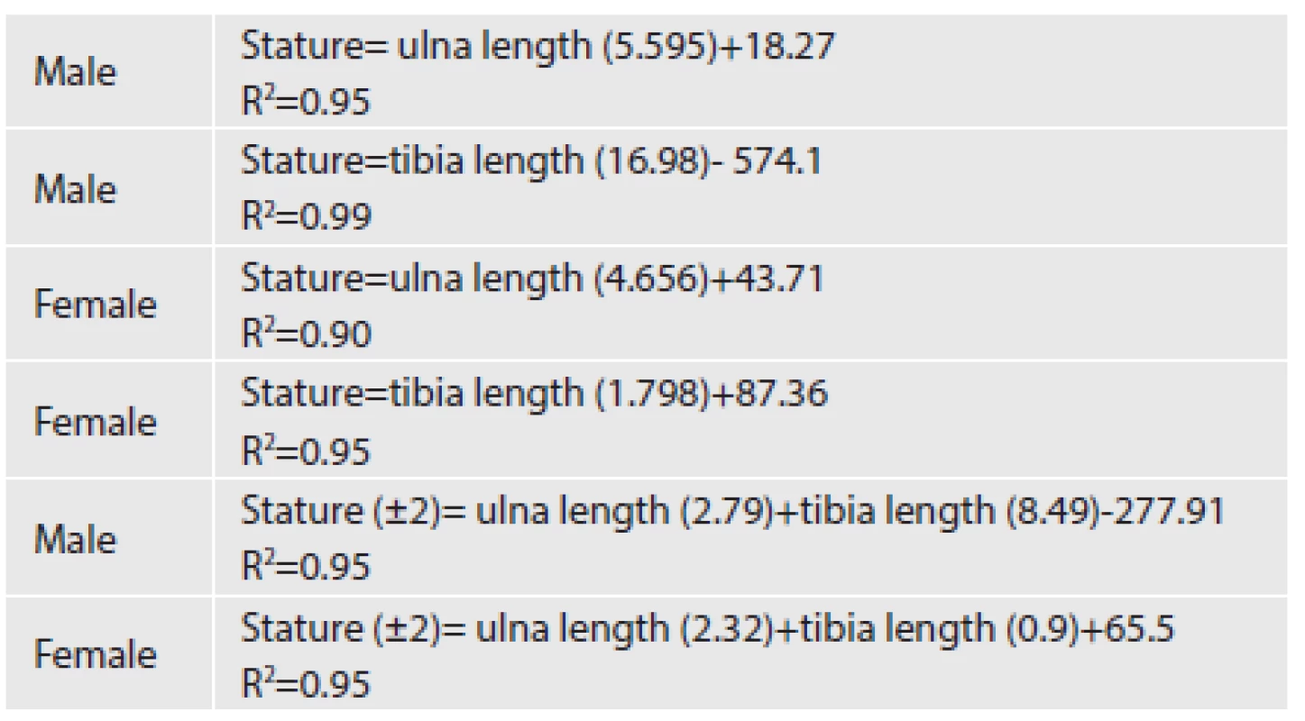
Do studie bylo zahrnuto celkem 49 dospělých mužů a 52 žen anatomicky zdravých náhodně vybraných ve dvou akademických centrech ve věkovém rozmezí mezi 20 a 40 lety. Výška postavy byla měřena naboso ve stoje. U každého jedince byl zhotoven rentgenový snímek pravého předloktí a pravého bérce v předozadní projekci. Na snímku byla měřena délka tibie a ulny. Byly sledovány demografické charakteristiky případů, včetně jejich věku a pohlaví. Vztah mezi vzrůstem a délkou loketní a holenní kosti jsou v publikaci znázorněny v diagramech a byly též definovány matematické vzorce.
Průměrná délka tibie u mužů byla 43.6 ± 0,4 cm a ulny 27.3 ± 0,6 cm. U žen délka tibie 40 ± 1.8 cm, ulny 25 ± 0,7 cm. Uvádíme grafy a regresní rovnice pro odhad výšky muže a ženy z délky loketní kosti.
Měření dlouhých kostí z RTG snímků při určování výšky postavy může být užitečné ve forenzní praxi při identifikaci osob. V předkládané studii byla měření prováděna na dospělých jedincích ve věkovém rozmezí 20-40 let v Teheránu, ale podobné studie v našem regionu by se měly zaměřit i na ostatní národnosti a věkové kategorie.
Klíčová slova:
stature – length of long bones – ulna – tibia – radiological evaluation – skeletal remains
Authors:
Mohammadreza Farsinejad; Samira Rasaneh 1; Nasim Zamani 2; Farkhondeh Jamshidi 3 4
Published in:
Soud Lék., 59, 2014, No. 2, p. 20-22
Category:
Original Article
Overview
Background:
We aimed to determine specific formulae by which we are able to estimate the body stature from the length of ulna and tibia calculated from the X-rays in order to be a reference for skeletal remains-based identification in Iranian population.
Methods:
The length of right ulna and tibia of 49 male and 52 female adults, who were anatomically healthy, were measured on the antero-posterior X-rays. Body height of each subject was also recorded.
Results:
Mean stature of the male and female adults was reported to be 171 ± 3.6 and 160 ± 3.9 centimeters (cm), respectively. Four single linear regression equations and 2 multiple regression equations were obtained.
Conclusions:
Lengths of ulna and tibia measured on the X-rays may be useful for estimation of the stature in cases of forensic personal identification.
Keywords:
stature – length of long bones – ulna – tibia – radiological evaluation – skeletal remains
Identity is a collection of the factors which help identifying a person from another. Medically, it consists of a series of general characteristics including sex, age, stature, and race (1). With respect to the racial, physical, and nutritional differences between different populations, it is essential to perform specific evaluations in each society in order to modify the previously existing references for determination of the identity (2). Estimation of the stature of the dead bodies using skeletal remains is one of the cornerstones of identification in forensic medicine which is possible to be performed by either anatomical or numerical measurement (3,4). In the anatomical method, the approximate stature of the body can be determined by direct measurement of the bones with few centimeters of difference. However, this method accompanies with two major difficulties. First, the total skeletal remains including the skull, bones of the lower extremities, and spinal column should be available while measuring the stature which is generally not possible, and second, the approximate thickness of the soft tissue of the head, calcaneous area, joint cartilages, and inter-vertebral disks should be added to the length of the bones measured which again is not usually accurate. In such a situation, a minimum of 6 to 8 centimeters (cm) of difference between the measured and true stature should generally be expected (3-5). In the numerical method, regarding the length of the upper or lower extremities and the stature, a mathematical formula is obtained which is possible to be generalized to the skeletal remains of the same population (6,7). Different studies have been performed in this regard in the western countries and United States, one of these is the Trotter’s and Gleser’s method (2). By application of their tables, stature is measured using the length of femur, tibia, and humerus bones and with respect to the sex and race (2,7,8).
Estimation of the stature based on the tibia height has previously been evaluated by Duyar and Pelin (9). They had concluded that when estimating height based on tibia length, the individual’s general stature category should be taken into consideration, and group-specific formulae should be used for short and tall subjects. Auerbach and Ruff also evaluated it in a group of North American populations and withdrew promising results (10). Since studies on this topic are few in our region, we decided to determine the relationship between the stature of the Iranian population and the length of the ulna and tibia using radiological evaluations in order to be a practical reference for identification of the Iranian population.
MATERIAL AND METHODS
A total of 49 male and 52 female anatomically healthy adults referring to Hazrat Rasoul Akram and Shohada Yaft Abad hospitals were randomly selected and included in the study. They had referred to these hospitals for problems except for orthopedic and bony structural disorders. Those with nutritional, musculoskeletal, congenital or acquired deformities, gonadal dysgenesia, and/or amputated right forearm and leg were not included. Their age range was between 20 and 40 years. After brief explanation and taking written consent forms as well as taking the approval from our regional ethics committee, their stature was measured barefoot and in standing position. For this purpose, the height between the vertex and the end point of calcaneous - while back of the shoulders and buttocks were touching the wall - was measured. Antero-posterior radiographs were taken from each case’s right forearm and leg. In the X-rays, length of the tibia was measured from the fossa between the internal and external intercondylar tubercles to the inferior articular surface of the tiba (Figure 1). Although the length of tibia is generally measured from lateral condyle to the medial malleolus, we did this because most of the skeletal remains of the large bones are eroded when discovered, especially in the prominent parts. We, therefore, selected non-prominent parts of the bone as landmarks for the measurements. Such a modification has already been tried by Vercellotti et al, as well (11). Length of the ulna was measured from the olecranon to the styloid process (Figure 1). For increasing the accuracy of the measurements, each measurement was repeated three times and the mean value of all the three times was used. Demographic characteristics of the cases including their age and sex were also recorded. The data was analyzed using Excel 2007 by application of scattered plots. With respect to the sex variable, the relationship between stature of the cases and length of their ulna and tibia were separately depicted in diagrams and mathematical formulae were obtained.

RESULTS
Mean stature of the males and females were 171 ± 3.6 centimeters (cm) and 160 ± 3.9 cm, respectively. Mean length of the tibia and ulna were 43.6 ± 0.4 and 27.3 ± 0.6 cm in men. These values were reported to be 40 ± 1.8 and 25 ± 0.7 cm in women.
The diagram of the estimation of the stature using the ulna in men and women has been shown in Figure 2. This was repeated for men and women for the tibia, as well (data not shown). The uni - or bi-variable obtained formulae showing the relationship between the stature and length of the bones have been exhibited in the Table 1.


DISCUSSION
In our country, despite the acceptable progression in different fields of forensic medicine, less attention has been paid to determination of identification using the body stature from the skeletal remains which is a very important essential point in identification. Nowadays, determining the body stature in Iran is being performed using the Trotter and Gleser tables which have been being used since the early 1900s in the black and white Americans (2,7). With respect to the affecting factors on the stature including the race, nutrition, and genetics as well as passing of about 110 years from the date of the generation of these formulae, it seems that their use is not only not helpful in determining the stature in the Iranian population, but also may cause incorrect identification in some situations. We, therefore, believe that using our formulae, determination of the stature may be more accurately performed. This is just the first step for future research which should improve determination of stature in our country. Out results are still not applicable to the practice as it was performed on a relatively small sample. Also, the method is applicable only for the age group 20-40 years and using the method in the older age groups will cause overestimation of the body height. Aware of all these limitations, we need to test our results on an independent sample.
CONCLUSION
Lengths of ulna and tibia measured on theX-rays may be useful for estimation of the stature in cases of forensic personal identification. Since the present study has been performed in the adult population (20 to 40 years) in Tehran, similar studies on other races and other age ranges are warranted in our region.
Correspondence address:
Nasim Zamani MD
Department of Clinical Toxicology,
Loghman Hakim Hospital,
Kargar Ave., Tehran, Iran
tel.: 00989122059290
e-mail: nasim.zamani@gmail.com
Sources
1. Cologlu AS. Adli Antropoloji Ders Notları. Istanbul Universitesi Adli Tip Enstitusu Yayini, Istanbul.1990.
2. Trotter M, Gleser GC. Estimation of stature from long bones of American Whites and Negroes. Am J Phys Anthropol 1952; 10 : 463-514.
3. Lundy JK. The mathematical versus anatomical methods of stature estimate from long bones. Am J Forensic Med Pathol 1985; 6 : 73-76.
4. Camps FE. Identification by the Skeletal Structures, Gradwohl’s Legal Medicine (3rd edn). Bristol: John Wright and Sons LTD; 1976 : 109-35.
5. Yayim Yili. Estimation of stature from tibial length. Journal of Forensic Medicine 1996; 12 : 87-93.
6. Jantz RL. Modification of the Trotter and Gleser female stature estimation formulae. J Forensic Sci 1992; 37 : 1230-1235.
7. Iscan MY. The Wisdom of Wilton Marion Krogman Fonder of Forensic Anthropology. Adli Tıp Dergisi 1990; 6 : 107-117.
8. Krogman WM. Iscan MY. The Human Skeleton in Forensic Medicine (2nd edn). Illinois: Thomas Publisher and Springfield; 1986 : 58.
9. Duyar I, Pelin C. Body height estimation based on tibia length in different stature groups. Am J Phys Anthropol 2003; 122 : 23-27.
10. Auerbach BM, Ruff CB. Stature estimation formulae for indigenous North American populations. Am J Phys Anthropol 2010; 141 : 190-207.
11. Vercellotti G, Agnew AM, Justus HM, Sciulli PW. Stature estimation in an early medieval (XI-XII c.) Polish population: testing the accuracy of regression equations in a bioarcheological sample. Am J Phys Anthropol 2009; 140 : 135-142.
Labels
Anatomical pathology Forensic medical examiner ToxicologyArticle was published in
Forensic Medicine

2014 Issue 2
-
All articles in this issue
- Ultrastruktural diagnosis of hypertrophic kardiomyopathy with β-aktin mutation in sudden death – case report
- Death Due to Perforation of Solitary Rectal Ulcer: Case Report
- Relationship between the stature and the length of long bones measured from the X-rays; modified trotter and gleser formulae in iranian population: A preliminary report
- Forensic Medicine
- Journal archive
- Current issue
- About the journal
Most read in this issue
- Death Due to Perforation of Solitary Rectal Ulcer: Case Report
- Ultrastruktural diagnosis of hypertrophic kardiomyopathy with β-aktin mutation in sudden death – case report
- Relationship between the stature and the length of long bones measured from the X-rays; modified trotter and gleser formulae in iranian population: A preliminary report
