-
Články
- Časopisy
- Kurzy
- Témy
- Kongresy
- Videa
- Podcasty
A new method of skin erythrosis evaluation in digital images
Autoři: P. Mezzana; T. Anniboletti; G. Curinga; M. G. Onesti
Působiště autorů: Department of Dermatology and Plastic Reconstructive Surgery, University of Rome “La Sapienza”, Italy
Vyšlo v časopise: ACTA CHIRURGIAE PLASTICAE, 49, 1, 2007, pp. 21-26
INTRODUCTION
The term color is used with different meanings in different technologies. To lamp engineers, color refers to a property of light sources. To graphic art engineers, color is a property of an object’s surface (under a given illumination). In each case, color must be physically measured in order to record and reproduce it.
The perception of color is a psychophysical phenomenon, and the measurement of color must be defined in such a way that the results accurately correlate with the visual sensation of color in a normal human observer. Colorimetryis the science and technology used to quantify and describe physically the human color perception.
Around 1930, Wright and Guild made independent visual experiments to derive color matching functions using three R/G/B (Red, Green, Blue) primaries, the results of which became the basis of the CIE colorimetry system.
RGB in graphics is both a way of specifying color and a way of viewing color. Graphics algorithms manipulate RGB colors, and the images produced by graphics algorithms are encoded as RGB pixels and displayed on devices that render these pixels by emitting RGB light.
Colored images are also used to specify color in graphics. These images may be captured by cameras or scanners, interactively drawn using tools such as Adobe PhotoShopTM, or algorithmically generated (1).
That color can be represented by three numbers, whether RGB or XYZ, is a direct result of the physiology of human vision. Electromagnetic radiation, with a wavelength in the visible range (370 to 730 nm), is converted by photo pigments in the retinal cones into three signals, which correspond to the response of the three types of cones. The cones convert this to three cone response values (L, M, S), that is, the cone sensitivities in the long, medium, and short wavelength regions defined by integrating the product of the spectral sensitivity curves and the incoming spectrum. Two important principles follow from this process:
- Trichromacy: all spectra can be reduced to precisely three values without loss of information with respect to the visual system.
- Metamerism: any spectra that create the same trichromatic response are indistinguishable. This means that two different spectra will look the same if they stimulate the same cone response. It is important at this point to distinguish between the perception of color and the creation of color. In practice, both can be described by three values, but the discussion up to this point has only covered perception. To use a digital color analogy, converting spectra to three cone response values corresponds to using a camera to create image pixels. We have not yet discussed how to display these pixels in a way that recreates the original sensation of color. The bridge to creation is provided by the color-matching experiments that underlie the CIE standards for measuring and specifying color. Using CIE colorimetry and a bit of linear algebra, the RGB color values used in computer graphics can be defined in a way that provides a direct link to perception. This creates a quantitative foundation for manipulating these values with respect to physical specifications of color such as color displays. It creates a link to scientific specifications of color, such as those created by modeling real surfaces and lights. It provides a way to integrate computer-generated imagery with color management systems, such as those used in the graphic arts. Finally, it provides an opportunity to integrate with current research in color appearance modeling, to provide renderings optimized for human color perception.
The major chromophobes for visible light in the epidermis are melanin and hemoglobin. The absorption and scattering property of the skin depends upon the path traversed by the light in the skin. The depth of penetration of optical radiation is dependent on the wavelength and on the physiologic state of the skin, such as in the erythema when the blood count of the subpapillary plexus increases and a greater amount of light is absorbed and less reflected (2–5). In order to provide an objective, accurate and quantitative color information about skin lesions, devices such as reflectance spectrophotometer and reflectance colorimeter have been successfully used during the past decade, though they are too expensive and technically complex to be handled in routine clinical situations (6–10). Spectroreflectometers measure the spectral reflectance of a test sample under a given geometrical condition, and most are calibrated by a reference standard traceable to a national metrology laboratory. Thus their measurement uncertainty first depends upon the uncertainty of the reference standard. Uncertainties also arise from the characteristics of the spectroreflectometer. Effects contributing to the uncertainty include wavelength error, detector nonlinearity, stray light, bandwidth, the geometrical conditions for both illumination and viewing, and measurement noise (11, 12). The RGB model is called “additive” because a combination of the three pure colors “adds up” to white light. When color images are captured by a digital camera, such as a CCD, the response of the three color bands is usually somewhat different from each other. For instance, one band may respond more strongly to intensity than another. If this is the case, then the resulting image will have a hue shift to that color when imaging a white or gray patch, e.g., if the blue channel is stronger than the red and green, the white patch will appear to have a bluish hue (13). Also, the responses of a CCD are generally nonlinear with respect to illumination. Many machine vision algorithms implicitly assume that camera response varies linearly with intensity, such as in edge detection that is based fundamentally on contrast between groups of pixels. We would like to calibrate the camera response in order to measure color from images objectively (14). The purpose of our study was to develop a standard system for computerized color image analysis of skin erythrosis modification after Intense Pulsed Light treatments, making it possible to compare readings taken by different observers in different environmental light conditions with the same digital photographic equipment.
MATERIALS AND METHODS
The trial was conducted over six months, between December 2004 and May 2005 at the Department of Dermatology and Plastic Surgery of University of Rome “La Sapienza”.
We evaluated 30 Caucasoid healthy subjects of both sexes, 18 females and 12 males, aged about 20 to 66 years old. The patients were chosen by their response to our parameters such as motivation to participate at the study, permission to take pictures, grade of skin erythrosis, informed consent. For the same reasons we excluded patients who did not satisfy the points above or who had previous treatments for skin erythrosis. The evaluation were conducted at room temperature (20 +/ - 4° C) and relative humidity varied between 40% and 60%. Before taking digital images, each volunteer was acclimatized under relax conditions for at least 30 min, with test site uncovered. Three points of standard colored paper (Red Green Blue) were applied with a plastic pattern using standard intersection lines in the involved area, and one point (White colored paper) in a non involved area for skin control (Fig. 1).
For 3 hours before the visit, the volunteer had not to smoke, drink coffee or alcohol, or take drugs. The pictures were taken using a Fujifilm FinePix S 20 PRO camera, in different light conditions, either pre than post Intense Pulsed Light (IPL) treatment, making it possible to compare readings taken by different observers in different environmental light conditions.
For every patient we took a series of pictures pre-treatment and after a 4/6 cycles of IPL.
The standard IPL treatment used was: 5–15 minutes, filter of 500–550 nm, double-triple impulse of 2.4–7 ms, delay 10–60 ms, repeated after 15–20 days, power respecting the device scale.
The images were obtained photographing the subjects with self-adhesive colored markers and some standard operative camera function (quality: fine; REC mode: normal; picture effect: normal; bright: segments and points 50/50; program: auto; macro and super macro: only for the details) except for the use of the flash and ambient light that were, on purpose, switched alternatively on and off for any patient to obtain the variability of above. The markers were circle-shaped adhesive tapes (each one of a diameter of 0.5 cm) (see Fig. 1). We put the color markers in the photographed skin area (except the white one): red above, green lateral below, blue medial below and a white point inserted in a non involved area (1 cm above the glabella) so as to place the device inside the erythematous skin area and the control in an area without erythrosis. These markers had not been photographed in standard illumination conditions. The resulting images were elaborated by Adobe Photoshop CS (Adobe Systems Incorporated© 1990–2003, version 8.1). The first elaboration was done in the acquired images before IPL treatment analyzing with the RGB system the color coefficient of the colored rings. Then was selected and indexed the cutaneous surface inside. The obtained data were inserted in a database to be correlated with the values obtained from the analysis of the images post treatment (24 hours after IPL treatment). The goal of our analysis was to evaluate objectively the grade of erythema. Erythema is a vasodilatation with increase of the blood flow red cell number. Hemoglobin is the principal cromophobo; it absorbs the green light and reflects the red light. An increase of hemoglobin is responsible for greater capture of green light and reflection of red light. The statistical analysis carried out on the pre-treatment images taken in different environmental conditions validated the reproducibility of the colorimetric measurement system. The relative colors of the red in different light conditions did not show any significant statistic change. In the next step the images obtained before the therapy with IPL were collected in a database and then compared with the post-treatment ones to verify the effective benefit. For every case was possible to verify the improvement grade obtained after IPL and quantify the results objectively and in numerical terms. The application of a white spot allowed us to eliminate almost all the errors due to the skin chromatic changes, etc. After quantifying the color value in RGB scale for any ring and surface, to obtain the value real red (R) it was necessary to subtract the value green (G). After quantifying the values of red of the “Ring” (Rc) and the “Area” (Ra) (Rc = Rring-Gring, Ra = Rarea-Garea) of the digital images in different light conditions, an analysis of normalization was performed in order to verify the effective reproducibility (Table 1, Fig. 2). To verify the accuracy of the system we effectuated a statistic analysis measuring the standard deviation. Validated the system of quantifying from digital images the values of Red, we have analyzed the real Red of images obtained post IPL treatment and normalized with the real red of digital images of pre treatment.
Fig. 1. Colored point in involved area 
Tab. 1. Digital images in different light conditions, an analysis of normalization 
Fig. 2. Digital images in different light conditions: a) light situation A, b) light situation B 
The values of the Real Red (normalized) were compared mathematically with the values obtained after the IPL treatment through this proportion: Ra:Rc (values of Red of pre-image) = X: R’c (values of R post treatment image) where X represents the RGB valor normalized to the referent image (R’a).
The system permits us to calculate the “real red image” from images post treatment, and by normalization process with the “real red” of pre-treatment images, it allows us to evaluate the efficacy of our treatment by IPL.
RESULTS
From a careful study on the pre-treatment digital images obtained in different environmental conditions we evaluated the grade of reproducibility of our procedure (see Table 1 and Fig. 2).
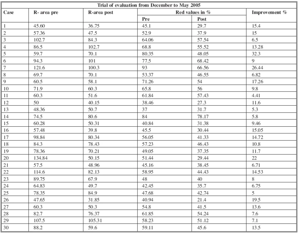
The statistic analysis of the standard deviation between the values of R obtained and the respective normalized valor normalized to the referent image did not show any significant statistical difference and allows us to achieve our goal: the reproducibility of the results. The method permits us to evaluate objectively the grade of improvement of the erythrosis after IPL therapy (Fig. 3 and Table 2).
Fig. 3. Objective evaluation of the grade of improvement of the erythrosis after IPL therapy: a) pre-treatment, b) post-treatment a)Before IPL treatment Control White point Adjacent skin RGB total 207.49 153.34 Control Real Red (Rc) 101.03 IMAGE Real Red (Ra) 45.60 b) Post 5 IPL treatment (after 4 months) Control White point Adjacent skin RGB total 215.34 173.02 Control Real Red (Rc) 123.41 IMAGE Real Red (Ra) 36.75 NORMALIZATION (POST on PRE) 36.75:123.41=X:101.03...X=30 IMAGE Real Red Pre 45.6:101.03=X:100 45.1% of the control IMAGE Real Red Post 30:101.03=X:100 29.7% of the control 15.4% improvement 
Tab. 2. Improvement in 30 cases after 5/6 IPL treatment 
DISCUSSION
Thanks to digital camera resolution improvements, nowadays it is possible to use it as a color measurement system.
The modern disposable software lets us analyze the digital images using color scales. We create a new useful method for the colorimetric exam of the skin.
The simplicity in the digital images acquisition and elaboration, the colorimetric analysis in numerical values of RGB scale of the cutaneous surfaces interested and the successive statistic elaboration allowed us to verify the reproducibility of the data.
Digital images in plastic surgery, dermatology and cosmetology represent an important element for the diagnostic and therapeutic evaluation of the pathologies under examination.
A simple electronic elaborator and a photographic analysis program permit us to quantify the grade of erythrosis of the cutaneous surfaces.
Our system provided the elaboration of the digital images and the following comparison with colored points (Red, Green, and Blue) to obtain determined colorimetric values in terms of comparison with fixed values (RGB).
The RGB color values used in computer graphics can be defined in a way that provides a direct link to perception. This creates a quantitative foundation for manipulating these values with respect to physical specifications of color such as color displays. It creates a link to scientific specifications of color such as those created by modeling real surfaces and lights. It provides a way to integrate computer-generated imagery with color management systems, such as those used in the graphic arts. It provides an opportunity to integrate with current research in color appearance modeling, to provide renderings optimized for human color perception.
Comparative analysis with the colors applied around the surface to be analyzed allows us to change the color value on a numerical scale (RGB scale) independently from the external light. In our study the colors R, G and B, assigned to the points applied on a determined surface, are not pure colors, due to the difficulty of finding them and ensuring the effective scientific value; instead, they are tonalities that allow us to compare in different light conditions and to measure objectively the grade of improvement of the erythrosis.
Comparing our results with the literature review, we validated the reproducibility of the system, confirming the study of Michele Setaro and Adele Sparavigna, DermIng, Institute of Clinical Research and Bioengineering, Monza (Milan), Italy, “Quantification of erythema using digital camera and computer-based color image analysis: a multicentre study”, adding some fundamental changes that allow the reproducibility of the system (15).
First, the device we have been using is not a marker that needs to be photographed in standard conditions (eliminating costs, difficulties and time spent), since the dependence of that system on the light, which represents the most difficult obstacle, can easily be bypassed with normalization. Our system makes it possible to analyze a bigger area where the device is placed, and adding a neutral control (see the white point), which can be considered a further value of comparison, allows us to obtain adjusted color measurements in different illumination conditions, in order to obtain data comparable between different researchers.
The white point is used to check the level of skin tan score. The RGB values of the white spot and the normal skin surrounding must not differ significantly between pre-treatment and post-treatment images.
The innovations introduced thanks to our study allow validating the system of colorimetric analysis of cutaneous surfaces as a very useful method to compare the erythrosis grade of the skin using a simplex statistic analysis of the obtained values on the comparison color.
The most relevant innovation out of the ones we introduced is the ability to calculate and assign a numerical value to a specific surface in RGB scale. The variable surface permitted the evaluation of the erythrosis grade before and after therapy.
We think that the further studies regarding this field will guarantee even more interesting results.
The evaluated surfaces in the RGB scale may be less and less dependent on variables, which increase the likelihood that errors will be made. The presence of wrinkles, unaligned superficial surfaces and skin imperfections, which change the camera focus, will be less relevant and easily avoided.
The natural marker R, G, B may provide on an absolute scale a definitive value to permit even broader evaluation in numerical and quantitative terms.
The accessibility of such a reliable, simple and cheap system to measure skin color could be of great benefit for the study of skin physiology, pathology, monitoring of treatments with a wide potential of application in dermatology and cosmetology, as well as for quantitative evaluation of the efficacy of therapies for skin lesions.
CONCLUSIONS
Digital image integration, cataloguing and elaboration by photographic programs allow us to define a new method of colorimetric analysis of cutaneous surfaces.
The possibility of tracing standard colorimetric values with a RGB system enables us to evaluate the erythema grade, color, independently from any other external variable.
The utility and potentiality of the system, and the ease of achieving the erythema grade pre and post treatment with IPL, will allow routine use of our method in the near future.
Address for correspondence:
Paolo Mezzana, MD.
Via Merulana 61/A
00185 Rome, Italy
E-mail: pmezzana@yahoo.it
Zdroje
1. Stone MC. Representing colors as three numbers. IEEE Comput. Graph. Appl., 25, 2005, p.78–85.
2. Porock D., Kristjanson L. Skin reactions during radiotherapy for breast cancer: the use and impact of topical agents and dressings. Eur. J. Cancer Care (Engl.), 8, 1999, p. 143–153.
3. Tucker SL., Turesson I., Thames HD. Evidence for individual differences in the radiosensitivity of human skin. Eur. J. Cancer, 28A, 1992, p. 1783–1791.
4. Turesson I. Characteristics of dose-response relationships for late radiation effects: an analysis of skin teleangiectasia and of head and neck morbidity. Radiother. Oncol., 20, 1991, p. 149–158.
5. Turesson I., Notter G. Skin reaction as a biologic parameter for control of different dose schedules and gap correction. Acta Radiol. Ther. Phys. Biol., 15, 1976, p. 162–176.
6. Pierard GE. EEMCO guidance for the assessment of skin color. J. Eur. Acad. Dermatol. Venereol., 10, 1998, p. 11.
7. Lévźque JL., Poelman MC., Legall F., De Rigal J. New experimental approach to measure the skin reflected light. Application to cutaneous erythema and blanching. Dermatologica, 170, 1985, p. 12–16.
8. Diffey BL., Oliver RJ., Farr PM. A portable instrument for quantifying erythema induced by ultraviolet radiation. Br. J. Dermatol., 111, 1984, p. 663–672.
9. Andreassi L., Casini L., Simoni S., et al. Measurement of cutaneous color and assessment of skin type. Photodermatol. Photoimmunol. Photomed., 7, 1990, p. 20–24.
10. Serup J., Agner T. Colorimetric quantification of erythema. A comparison of two colorimeters (Lange Micro Color and Minolta CR200) with clinical scoring scheme and laser Doppler flowmetry. Clin. Exp. Dermatol., 15, 1990, p. 267–272.
11. Lahti A., Kopola H., Harila A., Myllylää R., Hannuksela M. Assessment of skin erythema by eye. Laser Doppler Flowmeter, spectroradiometer, two-channel erythema-meter and Minolta chromameter. Arch. Dermatol. Res., 285, 1993, p. 278–282.
12. Karamfilov T., Weichold S., Kerstin K., Vilser W., Wollina U. Remittance spectroscopy mapping of human skin in vivo. Skin Res. Technol., 5, 1999, p. 49–52.
13. Takiwaki H., Shirai S., Kanno Y., Watanabe Y., Arase S. Quantification of erythema and pigmentation using a videomicroscope and a computer. Br. J. Dermatol., 131, 1994, p. 85–92.
14. Takiwaki H., Shirai S., Watanabe Y., Nagakawa K., Arase S. A rudimentary system for automatic discrimination among basic skin lesions on the basis of color analysis of video images. J. Am. Acad. Dermatol., 32, 1995, p. 600–604.
15. Setaro M., Sparavigna A. Quantification of erythema using digital camera and computer-based color image analysis: a multicentre study. Skin Res. Technol., 8, 2002, p. 84–88.
Štítky
Chirurgia plastická Ortopédia Popáleninová medicína Traumatológia
Článek ČESKÉ SOUHRNY
Článok vyšiel v časopiseActa chirurgiae plasticae
Najčítanejšie tento týždeň
2007 Číslo 1- Metamizol jako analgetikum první volby: kdy, pro koho, jak a proč?
- Kombinace metamizol/paracetamol v léčbě pooperační bolesti u zákroků v rámci jednodenní chirurgie
- Antidepresivní efekt kombinovaného analgetika tramadolu s paracetamolem
- Metamizol v terapii akutních bolestí hlavy
- Srovnání analgetické účinnosti metamizolu s ibuprofenem po extrakci třetí stoličky
-
Všetky články tohto čísla
- COMBINATION OF POSTERIOR INTEROSSEOUS AND HYPOGASTRIC FLAP FOR SKIN DEFECT RECONSTRUCTION IN HAND INJURIES
- BURIED UMBILICUS: AN IATROGENIC CAUSE OF A DISCHARGING UMBILICAL WOUND
- A new method of skin erythrosis evaluation in digital images
- ČESKÉ SOUHRNY
- NEO-PHALLOPLASTY WITH RE-INNERVATED LATISSIMUS DORSI FREE FLAP: A FUNCTIONAL STUDY OF A NOVEL TECHNIQUE
- AN OBJECTIVE EVALUATION OF CONTRACTION POWER OF NEO-PHALLUS RECONSTRUCTED WITH FREE RE-INNERVATED LD IN FEMALE-TO-MALE TRANSSEXUALS
- Acta chirurgiae plasticae
- Archív čísel
- Aktuálne číslo
- Informácie o časopise
Najčítanejšie v tomto čísle- NEO-PHALLOPLASTY WITH RE-INNERVATED LATISSIMUS DORSI FREE FLAP: A FUNCTIONAL STUDY OF A NOVEL TECHNIQUE
- AN OBJECTIVE EVALUATION OF CONTRACTION POWER OF NEO-PHALLUS RECONSTRUCTED WITH FREE RE-INNERVATED LD IN FEMALE-TO-MALE TRANSSEXUALS
- A new method of skin erythrosis evaluation in digital images
- COMBINATION OF POSTERIOR INTEROSSEOUS AND HYPOGASTRIC FLAP FOR SKIN DEFECT RECONSTRUCTION IN HAND INJURIES
Prihlásenie#ADS_BOTTOM_SCRIPTS#Zabudnuté hesloZadajte e-mailovú adresu, s ktorou ste vytvárali účet. Budú Vám na ňu zasielané informácie k nastaveniu nového hesla.
- Časopisy


