Synovial metaplasia of the endometrium: report of two new cases
Authors:
Michal Zámečník 1,2
Authors‘ workplace:
AGEL Laboratories a. s., Department of Pathology, Nový Jičín, Czech Republic
1; Medirex Group Academy n. o., Bratislava, Slovak Republic
2
Published in:
Čes.-slov. Patol., 55, 2019, No. 2, p. 112-113
Category:
Letters to Editor
To the Editor:
In 2015, Stewart and Leake described in the inflamed endometrium a peculiar lesion which they termed synovial-like metaplasia (SM) (1). They consider it similar to synovial-like metaplasia reported already in various extra-articular sites, such as tissue around prosthetic breast implants, adjacent to voice prosthesis, in the skin, around oral mucocele and in a lipoma (2-6). All their 11 cases were found in the endometrium with levonorgestel-releasing intrauterine device (L-IUS, Mirena). I would like to report two additional cases of this lesion, with immunohistochemistry of podoplanin that is a newly described marker for the synovial cells (7).
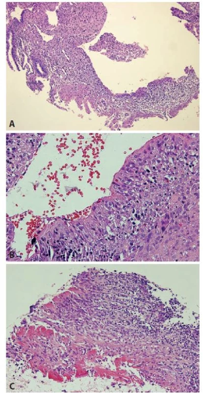
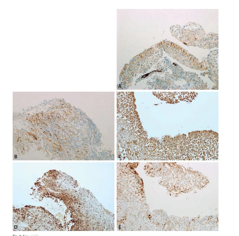
The female patients were 43-year-old para 2, gravida 2 (case 1), and 69-year-old para 1, gravida 1 (case 2), respectively. In both cases, the reason for dilatation, curettage and extraction of L-IUS was abnormal vaginal bleeding. Histological features (Fig. 1) in curettage specimens were very similar in both patients. Approximately 10 % of the endometrium showed SM of the mucosal surface, with morphology similar to that described by Stewart and Leake (1). Fibroblast-like spindle to ovoid cells were arranged perpendicular to the cavity. Focally, a fibrin material with polymorphonuclear leukocytes was seen on the surface. The endometrium showed mild chronic endometritis with some lymphoplasmacytic infiltrates, fibroblastic modulation and focal decuiduoid change of the stromal cells, and rare surface erosions. The endometrial glands were atrophic. Immunohistochemically, podoplanin (D2-40, Dako) was positive in approximately one third of the cells of SM, and it was focally seen in the stroma without SM (Figs. 2A and 2B) In addition, the cells of SM showed diffuse expression of both vimentin (3B4) and CD68 (KP1) (both from Dako) (Figs. 2C and 2D). Expression of CD10 (56C6, Zytomed) was found in 30 and 50 % of the cells of SM, respectively (Fig. 2E). The cells of SM were negative for pancytokeratin, epithelial membrane antigen, ER, PR, alpha-smooth muscle actin, desmin and h-caldesmon.


In the present cases, like in those of Stewart and Leaked, SM was found in the endometrium with L-IUS, and it was associated with known reactive/inflammatory changes caused by this device (8). It remains unclear why SM was never described in endometritis without L-IUS. Probably, a shearing or gliding effects of foreign body, which is absent in other types of endometritis, is a most important etiopathogenetic factor that plays a main role, whereas chronic inflammation and progestogen effect represent supporting factors. In all cases, the SM appeared to be only focal (seen always in less than 20% of the tissue), thus reflecting a mechanical influence of L-IUS on the nearby endometrium.
Regarding immunohistochemical features, our results were similar to those of Stewart and Leake. The cells were diffusely positive for vimentin and CD68, focally for CD10, and they lacked actin, estrogen receptor and progesterone receptor. Immunostain for podoplanin was not performed in the previous study (1). In our cases, podoplanin was expressed by numerous cells of the lesions. Podoplanin was found recently to be positive in the synovial cells, especially in the setting of chronic inflammation (7). Thus, its expression in our cases (along with positivity for CD68 and vimentin) can indicate ”true“ synovial differentiation/metaplasia of the stromal cells of the endometrium.
Correspondence address:
Michal Zamecnik, MD
Medicyt, s.r.o., lab. Trencin
Legionarska 28, 91171 Trencin
Slovak Republic
e-mail: zamecnikm@seznam.cz
tel.: +421-907-156629
Labels
Anatomical pathology Forensic medical examiner ToxicologyArticle was published in
Czecho-Slovak Pathology

2019 Issue 2
-
All articles in this issue
- Cytology of synovial fluid
- Cytopathology of soft tissue tumors
- Hormonal cytology
- Cytology of Ovarian cysts
- Potential use of immunohistochemistry for diagnosis precision in historic paraffin blocks
- Synovial metaplasia of the endometrium: report of two new cases
- Chronic dissecting aneurysm of ascending aorta with a large intramural thrombus and isolated aortic defects
- Czecho-Slovak Pathology
- Journal archive
- Current issue
- About the journal
Most read in this issue
- Hormonal cytology
- Cytology of synovial fluid
- Cytology of Ovarian cysts
- Cytopathology of soft tissue tumors
