-
Články
- Časopisy
- Kurzy
- Témy
- Kongresy
- Videa
- Podcasty
Inflammatory Monocytes and Neutrophils Are Licensed to Kill during Memory Responses
Immunological memory is a hallmark of B and T lymphocytes that have undergone a previous encounter with a given antigen. It is assumed that memory cells mediate better protection of the host upon re-infection because of improved effector functions such as antibody production, cytotoxic activity and cytokine secretion. In contrast to cells of the adaptive immune system, innate immune cells are believed to exhibit a comparable functional effector response each time the same pathogen is encountered. Here, using mice infected by the intracellular bacterium Listeria monocytogenes, we show that during a recall bacterial infection, the chemokine CCL3 secreted by memory CD8+ T cells drives drastic modifications of the functional properties of several populations of phagocytes. We found that inflammatory ly6C+ monocytes and neutrophils largely mediated memory CD8+ T cell bacteriocidal activity by producing increased levels of reactive oxygen species (ROS), augmenting the pH of their phagosomes and inducing antimicrobial autophagy. These events allowed an extremely rapid control of bacterial growth in vivo and accounted for protective immunity. Therefore, our results provide evidence that cytotoxic memory CD8+ T cells can license distinct antimicrobial effector mechanisms of innate cells to efficiently clear pathogens.
Published in the journal: Inflammatory Monocytes and Neutrophils Are Licensed to Kill during Memory Responses. PLoS Pathog 7(12): e32767. doi:10.1371/journal.ppat.1002457
Category: Research Article
doi: https://doi.org/10.1371/journal.ppat.1002457Summary
Immunological memory is a hallmark of B and T lymphocytes that have undergone a previous encounter with a given antigen. It is assumed that memory cells mediate better protection of the host upon re-infection because of improved effector functions such as antibody production, cytotoxic activity and cytokine secretion. In contrast to cells of the adaptive immune system, innate immune cells are believed to exhibit a comparable functional effector response each time the same pathogen is encountered. Here, using mice infected by the intracellular bacterium Listeria monocytogenes, we show that during a recall bacterial infection, the chemokine CCL3 secreted by memory CD8+ T cells drives drastic modifications of the functional properties of several populations of phagocytes. We found that inflammatory ly6C+ monocytes and neutrophils largely mediated memory CD8+ T cell bacteriocidal activity by producing increased levels of reactive oxygen species (ROS), augmenting the pH of their phagosomes and inducing antimicrobial autophagy. These events allowed an extremely rapid control of bacterial growth in vivo and accounted for protective immunity. Therefore, our results provide evidence that cytotoxic memory CD8+ T cells can license distinct antimicrobial effector mechanisms of innate cells to efficiently clear pathogens.
Introduction
Immunological memory is a central feature of the adaptive immune system and relies on the maintenance of antigen-experienced B cells and T cells. In general, a memory immune response is faster and stronger than a primary response because of the increased frequency, improved functional characteristics, and preferential localization of the memory cells in peripheral tissues [1]–[2]. Memory CD8+ T cells represent one major effector arm of the adaptive immune system that maintains long-lived protective immunity against intracellular bacteria, protozoa, and viruses [1]. Although CD8+ T cells are mainly appreciated for their capacity to kill infected cells, they can express various effector mechanisms that potentially contribute to host defense against infections [3]. Former reports have suggested that the antimicrobial potential of CD8+ T lymphocytes is also reflected in their ability to rapidly produce inflammatory cytokines such as IFN-γ, TNF-α and CCL3 that promote the control of the growth of intracellular pathogens such as Francisella tularensis, Leishmania major or Listeria monocytogenes (Lm) by phagocytes [4]–[6]. In the case of a memory response, both the increased numbers of recruited phagocytes and their faster or chronic activation allow for an optimized clearance of intracellular pathogens [6]–[9]. This view is based on the assumption that phagocytes usually exhibit a qualitatively and quantitatively comparable response each time the same pathogen is encountered. However, it is not known whether and how the antimicrobial activities of innate phagocytes that are expressed during a primary immune response are modulated during recall infections.
Phagocytes such as macrophages, neutrophils and monocytes play a critical role during primary infections to fight intracellular pathogens. Both neutrophils and macrophages are well-established as important cellular effectors of innate immune defense, and it is clear that circulating monocytes also contribute significantly to the defense against a range of microbial pathogens [10]–[11]. Phagocytic cells recognize and engulf pathogens by expressing specific receptors of microbial products (pattern recognition receptors, PRRs), of the complement system or of antibodies complexed to pathogen-derived molecules [12]. Following microbial internalization, early phagosomes undergo numerous steps of maturation that are concomitant with important changes of their associated proteins and luminal pH acidification. Acidification results from the fusion of phagosomes with pre-existing lysosomes, leading to the activation of hydrolytic enzymes which act optimally at acidic pH (4.5–5.0) to enable the degradation of micro-organisms [13]. In addition to acidification, another critical mechanism involves recruitment to the phagosomal membranes of the nicotinamide adenine dinucleotide phosphate (NADPH) oxidase complex subunits which in turn generate the reactive superoxide ions that accumulate in the phagosomal lumen [14]. Superoxide is further converted to several highly toxic radical oxygen species (ROS) that degrade phagosomal content and facilitate pathogen killing [15]–[16]. Generation of ROS also induces phagosomal pH increase which subsequently activates neutral proteases that digest and kill microorganisms. Upon activation, phagocytes generate other important antimicrobial effector molecules such as reactive nitrogen species (RNS) produced by the inducible nitric oxid synthase (iNOS) that interact with ROS to exert very toxic effects on intraphagosomal engulfed microorganisms [17]. Collectively, these antimicrobicidal effectors activities provide a toxic phagosomal environment that efficiently limits pathogen proliferation. However, some pathogens including bacteria such as Listeria, Shigella, Rickettsia and group A Streptococcus (GAS) have evolved strategies to quickly escape from this hostile phagosomal environment to the more hospitable milieu of the host cell cytosol [18]. After escape into the cytosol, some of these bacteria can be trapped in large cytosolic vacuoles and ultimately be degraded within autolysosomal compartments [19]–[21]. This process, known as autophagy, has been more recently described as an intrinsic host defense mechanism for recognition and elimination of intracellular cytosolic or vacuolar pathogens [19].
In a previous study using mice intravenously inoculated with Lm as an infection model, we have shown that innate phagocytes can be activated by the Macrophage Inflammatory Protein 1α (MIP-1α/CCL3) that is secreted by memory CD8+ T cells upon antigen-driven reactivation [6]. CCL3 induced a rapid TNF-α secretion by innate inflammatory monocytes, which further promoted the production of ROS by both monocytes and neutrophils. We also found that ROS generation depended on CCL3 and was required for killing of Lm. In the present work, taking advantage of the same experimental settings in which the vast majority of bacteria were found inside phagocytic cells of the spleen [22]–[23], we investigated the modulation of the antimicrobial effector activities expressed by innate phagocytes in the course of the secondary infection and the impact this have on the clearance of the bacteria from infected tissues.
Results
A major role for CCL3-mediated reactive oxygen species (ROS)-dependent killing of bacteria in protection of immunized mice
CD8+ T cells usually protect against intracellular microbes by the recognition and the lysis of infected cells, via a perforin and/or a Fas/Fas-ligand-mediated mechanism of cytolysis. We recently demonstrated in the mouse Lm infection model that memory CD8+ T cells are also able to protect by orchestrating the activation of effector cells of the innate immune system via the release of the chemokine CCL3/MIP1-α [6]. Indeed, the transfer of CCL3-deficient effector [24] or memory [6] CD8+ T cells to infected mice does not confer protective immunity to these mice. Moreover, the protection achieved by wt memory CD8+ T cells infused into CCL3−/− recipient mice is totally abrogated upon CCL3 neutralization [6]. At least 2 recent reports have documented that memory CD8+ T cells induced in a bacterial and a viral infection indeed can produce high levels of CCL3 and that expression of this gene is enhanced in highly differentiated, long-lived memory cells that can confer immunological protection [25]–[26]. We further compared this mechanism to the classical CD8+ T cell-dependent effector mechanisms. For this, wt or perforin-deficient mice were immunized by infection with Lm (then referred to as “memory mice”), and 3 weeks later some animals were treated with anti-CCL3 serum or anti-IFN-γ neutralizing mAb before the challenge infection (Figure 1A). While animals lacking perforin exhibited only a modest increase in susceptibility (∼10 fold) compared to wt memory mice [27], none of the anti-CCL3-treated groups controlled the challenge infection. These mice had more than 106 bacteria in the spleen, a number close to that found in primary and in memory p47phox−/− infected mice that lack functional NADPH oxidase complexes and cannot produce ROS (Figure 1B). Interestingly, when memory mice were treated with a neutralizing Ab against IFN-γ, an important immunomodulatory cytokine of effector and memory CD8+ T cells, only about a 10 fold loss of protection was observed in wt mice (Figure 1A) [28]. Thus, whereas perforin - and IFN-γ accounted for a significant degree of protection in wt memory animals, the current results revealed a substantially greater role for CCL3 and ROS in protective memory responses to Lm.
Fig. 1. CCL3/ROS+-mediated killing of bacteria is a major mechanism of protection in memory mice. 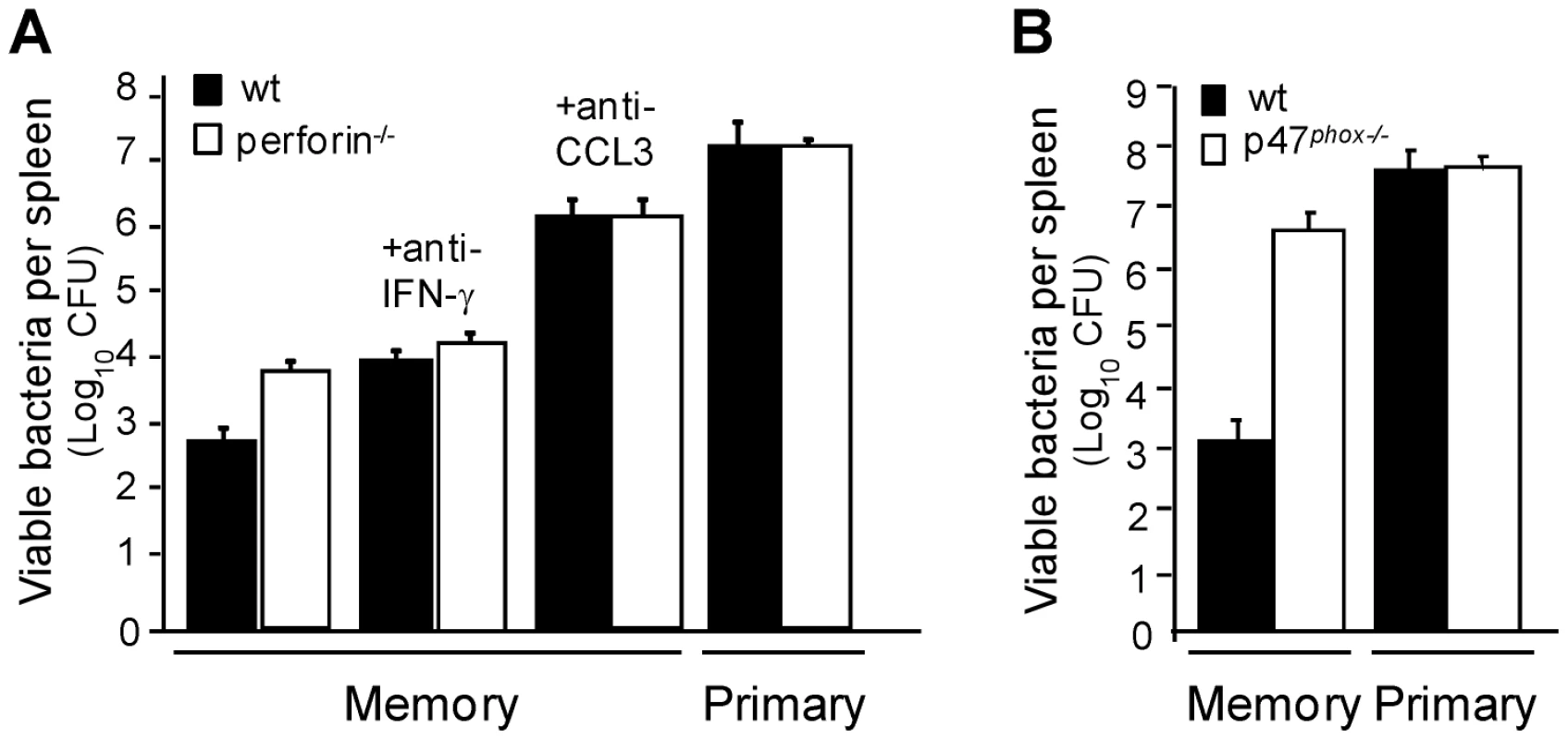
Wt (black bars) (A–B) and perforin−/− mice (white bars) (A) or p47phox−/− mice (B) were injected with PBS or immunized with Lm. One month later mice were treated or not with anti-CCL3 serum or anti-IFN-γ mAb and subsequently challenged with 3×10⁵ wt Lm for 2 days. Data show the number of bacteria CFUs (mean±SE) in a pool of two independent experiments with n = 6–9 mice per group. Improved antimicrobial vacuolar effector functions in phagocytes during memory responses
To study the modulation of the effector functions of phagocytes during a memory response, we first defined the cytofluorometry gating strategies that allowed us to identify the major innate phagocyte populations of the spleen. While neutrophils (CD11bhighF4/80lowLy-6Ghigh) and macrophages (CD11bmedF4/80highLy-6Gmed) are well-defined in the literature (Figure 2A), blood-monocytes that enter inflamed tissues and express antimicrobial functions have been variously described as inflammatory monocytes, [29], TNF-α/iNOS producing-dendritic cells (Tip-DCs) [30] or mononuclear phagocytic cells (MPCs) [6] depending on the experimental context. Since all of these cells exhibit a comparable cell-surface phenotype (CD11bmed/highF4/80medLy-6GlowCX3CR1lowCD11clowLy-6Chigh), produce TNF-α, RNS and ROS (Figure 2A, S1), it is likely that inflammatory monocytes, Tip-DCs and MPCs are the same cell population, which we refer to as inflammatory and/or ly6C+ monocytes.
Fig. 2. Increased frequencies of ROS+ inflammatory monocytes and neutrophils that generate higher levels of ROS during the secondary infection. 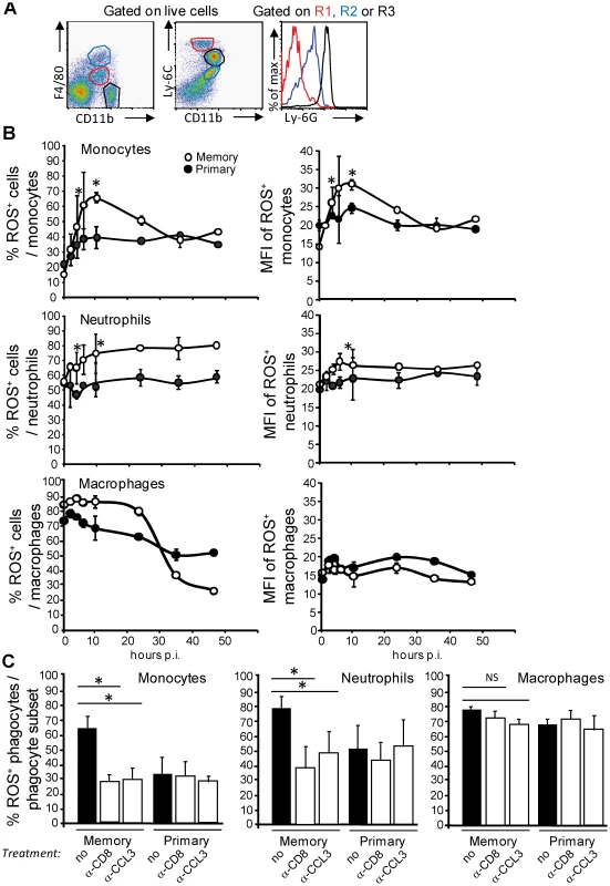
(A) Spleen cells from wt mice (3/group) were independently analyzed by FACS for surface expression of F4/80, CD11b, Ly-6C and Ly-6G. Data show representative FACS profiles in a representative (out of 2) experiment. Red gate: inflammatory Ly6C+ monocytes, black gate: neutrophils, blue gate: macrophages. (B) Mice (9–10 per group) primarily injected with PBS (closed circles, primary) or 0.1xLD50 (3×10³) wt Lm (open circles, secondary) were challenged 30 days later with 10xLD50 (3×10⁵) wt Lm. At the indicated times after challenge, spleen cells were restimulated with Heat Killed Lm (HKLM) in the presence of hydroethidine and analyzed by FACS for CD11b and Ly-6C expression. Data show the number (mean +/− SE) (left panels) and the MFI (mean +/− SE) (right panels) of ROS-producing inflammatory monocytes (upper panel), neutrophils (middle panel) and macrophages (bottom panel) per total gated inflammatory monocytes, neutrophils or macrophages and are representative of a pool of 2–3 replicate experiments. (C) Primary and memory mice (10/group) were treated 30 days later with a control isotype or serum (black bars), anti-CD8 or anti-CCL3 (white bars) and challenged with 3×10⁵ wt Lm. At 10 hrs after the infection, the frequencies of ROS-producing cells were analyzed as in (B). Data result from the pool of 2 independent experiments. P (*<0.05) values were calculated in (B–C) with n = 9–10. In addition to the generation of ROS, phagocytic cells also express other important antimicrobial activities within their phagosomes during activation such as the generation of reactive nitric species (RNS) and the rapid modulation of the pH of the phagosomal lumen [31]. Since the enzymes that produce NO (the inducible nitric oxide synthase, iNOS) and ROS (the NADPH oxidase) can be activated by the release of IFN-γ and TNF-α, and because levels of these cytokines are much higher during the memory response, we monitored the activation of both enzymatic complexes during a primary as compared to a memory immune response. Mice injected with PBS or with Lm were challenged a month later with Lm (primary and memory responses, respectively), and the frequencies and mean fluorescence intensities (MFI) of ROS-producing and iNOS-expressing cells were measured by flow cytometry (Figure 2B–C, S2 and S3). The numbers and frequencies of ROS-producing inflammatory monocytes and neutrophils measured during a memory response (open symbols) were 1.3 to 2.4 higher than those of a primary response (closed symbols) (Figure 2B left; 2C, upper and middle panels and S2). In addition, the MFI of ROS+ ly6C+ monocytes was 1.2 to 1.5 fold increased compared to that of the ROS+ neutrophils and macrophages –at least the population of macrophages that we could recover from the spleen, suggesting that ly6C+ monocytes were intrinsically able to produce higher level of ROS than other effector phagocytes (Figure 2B, right panels). Such augmented effector activities depended on memory CD8+ T cells and CCL3 since immune mice treated either with an anti-CD8 depleting mAb or an anti-CCL3 neutralizing serum exhibited a lower frequency (∼30–40% instead of 60–70%) of ROS-producing inflammatory monocytes or neutrophils after challenge (white bars), equivalent to that found in primary infected animals (white and black bars) (Figure 2C, upper and middle panels).
Of note, while the subset of macrophages recovered from the spleen accounted for the large majority of ROS-producing phagocytes at steady state (data not shown), frequencies, numbers and MFI of these ROS+ macrophages did not vary neither during the primary nor the memory response (Figure 2B–C bottom panels and S2). Interestingly, whereas memory CD8+ T cells can promote ROS production by phagocytes, the frequencies of iNOS-expressing cells were similar in primary (closed symbols) and memory (open symbols) infected mice (Figure S3A). Moreover, in contrast to oxidative burst-deficient mice (Figure 1B & [6]), memory mice lacking the iNOS enzyme are equivalently protected during the secondary challenge as wt animals (white bars) and exhibited ∼5,000–6,500 fold less bacteria in the spleen than primary infected wt or iNOS−/− mice (black bars) (Figure S3B). Therefore, memory CD8+ T cells allow (i) increased numbers of inflammatory monocytes and neutrophils to be activated and (ii) the differentiation of effector monocytes and neutrophils that produce higher quantities of ROS on a per cell basis.
While an acidic luminal pH is essential for optimal activity of numerous microbicidal agents in macrophages and neutrophils, the NADPH-oxidase complex may also mediates the increase of the pH in phagocytic vacuoles that could lead under some experimental conditions to the activation of neutral proteases that kill microorganisms [14], [32]–[33]. Since phagocytes exhibited an increased production of ROS during a memory response, we monitored the phagosomal pH inside ly6C+ monocytes, neutrophils and macrophages from primary and memory mice 6 hrs after the infection (Figure 3, S4 and not shown). For this, latex beads were coated with pH-sensitive and pH-insensitive fluorescent dyes and incubated with splenocytes for 20 minutes to allow uptake of beads by phagocytes. Cells were further stained for the expression of different cell surface markers expressed by phagocytes and the fluorescence intensity of the phagocytosed beads was quantified by FACS. The ratio in fluorescence intensity between the two dyes directly reflected the pH inside the phagosomes as calibrated on a standard curve [34] (Figure S4A). We found that inflammatory monocytes which produced the highest levels of ROS per cell (Figure 2B, left panel), exhibited increased pH (∼5.2) inside their phagosomes in memory as compared to those of primary infected mice (∼4.45) (Figure 3, left panel). As expected, the intra-phagosomal pH for neutrophils [32]–[33], was also augmented in memory (∼5.2) versus primary (∼4.6) infected animals (Figure 3, right panel) [32]–[33]. These results were in sharp contrast with the intra-phagosomal pH of the splenic macrophage subset, which remained acidic (Figure S4B).
Fig. 3. The phagosomal pH inside inflammatory monocytes and neutrophils rises during a secondary immune response. 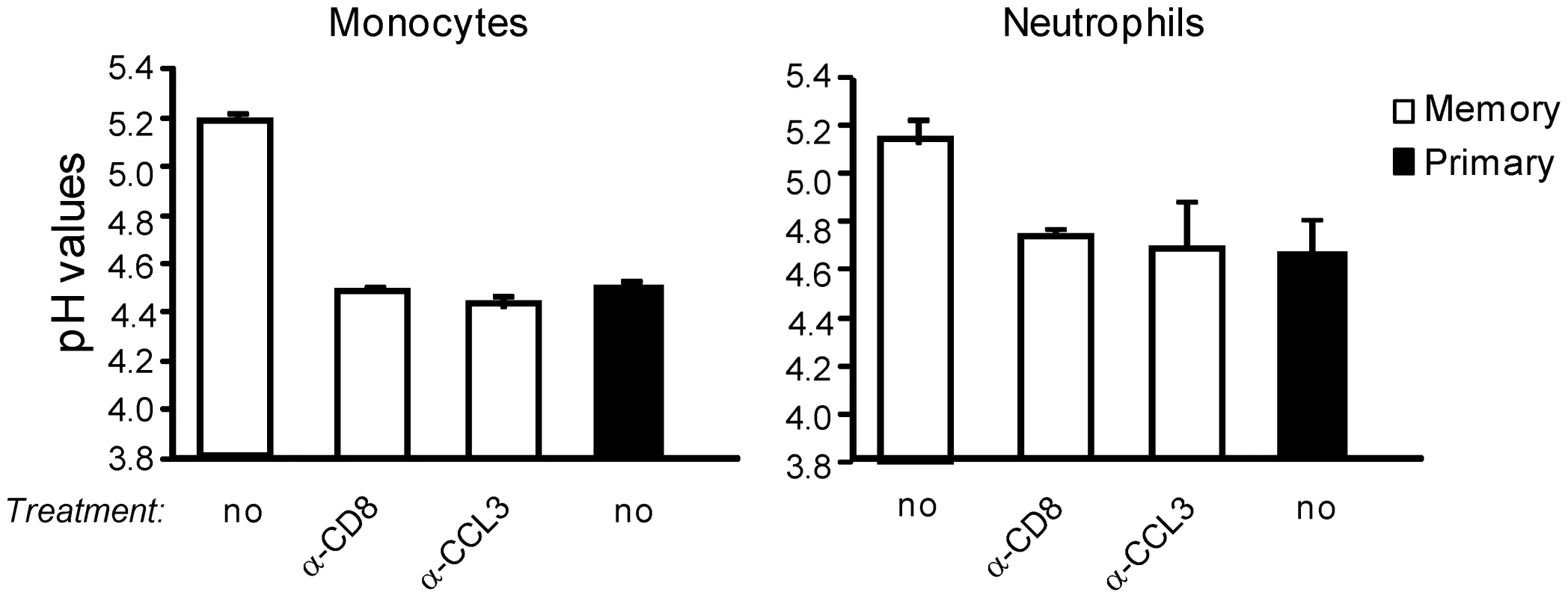
Primary (black bars) and memory mice (2–3/group) treated or not with anti-CD8 mAb or anti-CCL3 serum (white bars) were challenged with 3×10⁵ wt Lm for 6 hrs. Spleen cells were then incubated with latex beads coupled with pH sensitive and insensitive fluorochromes, stained for surface expression of Ly-6C and CD11b, and analyzed by FACS. Data show the pH values calculated for inflammatory monocytes and neutrophils in one (out of 2) independent experiments. Similar acidic pH values that reached pH ∼4.5 three hrs after phagocytosis have also been observed for the macrophage cell lines RAW264.7 and J774 in vitro under similar experimental conditions [35]. The higher pH of the phagosomes of inflammatory monocytes and neutrophils from infected memory mice depended on CCL3+ memory CD8+ T cells, since treatment of memory mice with an anti-CD8 depleting mAb or an anti-CCL3 neutralizing serum induced a pH drop equivalent to that of primary infected mice. Collectively, our results show that during a memory response, innate cellular effectors -inflammatory monocytes and neutrophils - exhibit an inhospitable environment inside their phagosomes that helps better clearance of bacteria and likely is more toxic for pathogens than in the course of a primary response.
Efficient prevention of cytosolic escape of Lm in phagocytes from immunized mice
During Lm infection, the initial step of escape from the primary vacuole of phagocytosis to the cytosol of infected host cells is critical for bacterial survival [36]–[37]. Since Lm is rapidly trapped in such vacuoles [38], we reasoned that the most efficient way of controlling Lm growth is to prevent its escape to the cytosol of infected phagocytes. If this hypothesis is true, Lm multiplication between primary and memory infected animals would be expected to differ as early as during the first few hrs of infection. To investigate this possibility, we monitored Lm multiplication inside the spleen of primary and memory challenged animals that were concomitantly treated with an anti-CD8 depleting Ab, anti-CCL3 neutralizing serum or control goat serum (Figure 4 and not shown). As expected, primary or memory infected mice treated with anti-CD8 or anti-CCL3 did not control the infection. These mice exhibited 6.6 and 20 times more bacteria in the spleen respectively 9 and 12 hrs after the challenge infection than memory mice either untreated (white circles, plain line) or injected with control isotype matched Ab or serum (not shown). At 24 hrs, between 23 and 216 times more bacteria were measured in the groups of mice that did not control the infection (Figure 4A). Therefore, memory mice (open symbols) have already cleared 40%, 72% and 85% of the bacteria colony forming units (CFUs) 1.5, 6 and 9 hrs after the challenge infection as compared to primary infected animals (closed symbols) (Figure 4B). This result showed that Lm killing began very early after challenge of memory mice. Because several effector activities of phagocytes were improved at these time points, we hypothesized that killing of Lm could happen inside the primary vacuoles of phagocytes and with a higher efficacy in mice with memory responses.
Fig. 4. CCL3+ memory CD8+ T cells control bacterial growth during a secondary infection through a vacuolar and a cytosolic mechanism. 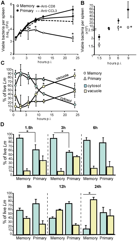
(A–B) Primary and memory wt mice (10/group) were treated (A) or not (B) with anti-CD8 or anti-CCL3, and challenged with 3×10⁵ wt Lm. Mice were sacrificed at the indicated times after the infection. (A–B) show the number of bacteria CFUs (mean +/− SE) in the spleen in a pool of 2 replicate experiments. P values were calculated between groups of mice injected with PBS or immunized with wt Lm (n = 10). (C–D) Primary (triangles) and memory mice (rounds) (9–12/group) were challenged one month later with 3×10⁵ wt-L029 Lm. At the indicated times after infection, spleen cells treated or not with chloramphenicol, lysed and plated. (C) Graphs show the frequencies (mean +/− SE) of cytosolic (blue) and vacuolar (yellow) Lm CFUs in phagocytes in a pool of 3–4 experiments (n = 9). (D) is another representation of these data with histograms reporting frequencies (mean +/− SE) of cytosolic (blue bars) and vacuolar (yellow bars) Lm CFUs. To determine whether the early killing of Lm indeed takes place inside such vacuoles, we took advantage of a recombinant Lm (wt-L029) which expresses an antibiotic resistance to chloramphenicol upon reaching the cytoplasm of infected cells [39]. Splenocytes were incubated with chloramphenicol for a short period of time, which kills extracellular wt-L029 Lm and those that are inside vacuoles, only allowing survival of Lm that escaped to the cytosol (Figure S5). Primary and memory mice were challenged with wt-L029 Lm and the frequencies of vacuolar (chloramphenicol-sensitive) and cytosolic (chloramphenicol-resistant) viable bacteria inside phagocytes were determined 1.5, 3 and 6 hrs post-infection (Figure 4C–D). In primary infected mice, 60% of viable Lm localized to the cytosol and 40% to the vacuoles of cells 1.5 and 3 hrs later (Figure 4D). In contrast, while only 5–10% of the viable bacteria were found in the vacuoles of the phagocytes from memory mice (yellow bars), the vast majority (∼90–95%) were recovered from the cytosol (blue bars). At 6 hrs, bacterial localization was similar between primary and memory mice, with ∼90% of viable Lm localized to the cytosol and ∼10% to the vacuoles of phagocytes. Because the large majority of viable bacteria were recovered from the cytoplasm of infected cells in memory mice, these findings suggested that Lm was killed with a higher efficacy in the vacuoles of phagocytes from memory compared to primary infected mice. To provide further support to this hypothesis, we determined the absolute numbers of viable bacteria inside the vacuoles of phagocytes from primary and memory mice 1.5 hrs after the infection with wt-L029 Lm (Figure 5A). At this early time point following challenge, memory mice already exhibited ∼30–50% fewer bacteria in the spleen than primary infected mice. Moreover, memory mice exhibited two fold less viable bacteria in the vacuoles than animals with primary infections. To formally demonstrate the improved killing of Lm inside the vacuoles of phagocytes during the memory response, primary and memory mice were challenged with listeriolysin (LLO)-deficient (ΔLLO) Lm that remained trapped inside the vacuoles, and the number of viable Lm was determined 1.5 hrs later. As for wt Lm, memory mice infected with ΔLLO-Lm exhibited 2.5 times less viable bacteria than primary infected animals (Figure 5B).
Fig. 5. Early killing of Lm requires ROI and takes place inside the vacuoles of phagocytes from secondary infected mice. 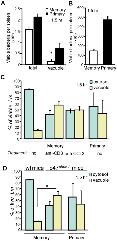
Primary and memory mice (9–12/group) were challenged one month later with 3×10⁵ wt-L029 Lm unless otherwise indicated. 1.5 hr post-infection, spleen cells treated or not with chloramphenicol, lysed and plated. (A) shows the number (mean +/− SE) of Lm CFUs recovered from the spleen. In (B), primary and immune mice (4–5/group) were challenged with 2×10⁸ ΔLLO Lm. The number of Lm CFUs (mean +/− SE) per spleen is shown in a pool of 2 experiments. In (C), primary and memory mice (4–6/group) were treated or not with anti-CD8 or anti-CCL3. Histograms show the frequencies (mean +/− SE) of cytosolic (blue bars) and vacuolar (yellow bars) Lm CFUs in phagocytes in a pool of 2 experiments. In (D), primary and memory p47phox−/− and wt C57BL/6 mice (5–6/group) were challenged with 3×10⁵ wt-L029 Lm. Histograms show the frequencies (mean +/− SE) of cytosolic (blue bars) and vacuolar (yellow bars) Lm CFUs in phagocytes in a pool of 2 experiments (n = 5). To assess whether the improved killing of Lm occurring inside the vacuoles of the phagocytes resulted from the enhanced activity of the NADPH-oxidase complex during the memory response, primary or memory mice lacking the inducible p47phox subunit of the NADPH-oxidase complex, or wt mice treated with either anti-CD8, anti-CCL3 or control Abs were infected with wt-L029 Lm (Figure 5C–D). p47phox−/− and wt memory mice treated with the anti-CD8 or the anti-CCL3 exhibited similar frequencies (∼40–60%) of viable vacuolar Lm as primary infected wt and p47phox−/− mice. Thus, taken together our results suggested that during a memory response, phagocytes are conditioned by the memory CD8+ T cells to generate a more effective bacteria-killing environment inside their early vacuoles of phagocytosis, and that this process is directly dependent upon increased activity of the NADPH-oxidase complex.
Engulfment of cytosolic Lm in vacuolar structures of phagocytes from previously immunized mice
While at very early time points after the infection (0–3 hrs), Lm was most efficiently killed within vacuoles of phagocytes from memory mice, the percentage of viable vacuolar bacteria (∼85%) and therefore the rate of phagosomal killing was comparable 6 hrs after the infection in primary and memory infected mice (Figure 4D). However, despite equivalent phagosomal elimination of Lm, the number of viable bacteria was significantly diminished in memory as compared to primary infected animals (Figure 4A–B). We therefore hypothesized that this either resulted from the more efficient early phagosomal killing of Lm or from other killing mechanisms distinct from those expressed inside the primary phagocytic vacuoles. If the first hypothesis were to be true, we expected to find a comparable distribution of the viable bacteria between the 2 groups of mice. We thus investigated the subcellular localization of Lm in infected splenocytes from primary and memory mice challenged with wt-L029 Lm (Figure 4C–D). Between 9 and 24 hrs after the infection, the relative proportion of viable cytosolic bacteria inside the phagocytes from memory mice drastically decreased from ∼60% to 15% compared to that of primary infected animals which only decreased about 20%. Viable Lm inside the cytosol of infected cells (i.e., chloramphenicol resistant) were rapidly lost and further recovered from vacuolar structures (i.e., chloramphenicol sensitive), suggesting that bacteria which had escaped to the cytosol of infected cells were rapidly re-engulfed into vacuoles. We reasoned that this could either result from Lm spreading from one cell to another or from the reinternalization of the free cytosolic Lm into phagosomal structures inside the same infected cell. To discriminate between the two possibilities, we used Lm lacking ActA that neither can spread from cell to cell nor infect neighboring cells. For that purpose, we generated an ActA-deficient L029 Lm strain that expressed the chloramphenicol resistance gene when reaching the cytosol of infected cells (Figure 6A). Upon inoculation of wt or ActA-deficient L029 Lm, phagocytes from primary and memory mice both exhibited similar frequencies of vacuolar viable bacteria, suggesting that the appearance of such vacuole-containing bacteria did not result from Lm spreading to neighboring cells but rather from Lm reinternalization inside the same infected cell.
Fig. 6. Cytosolic bacteria are engulfed and killed via ROI in the vacuoles of phagocytes during a protective secondary response. 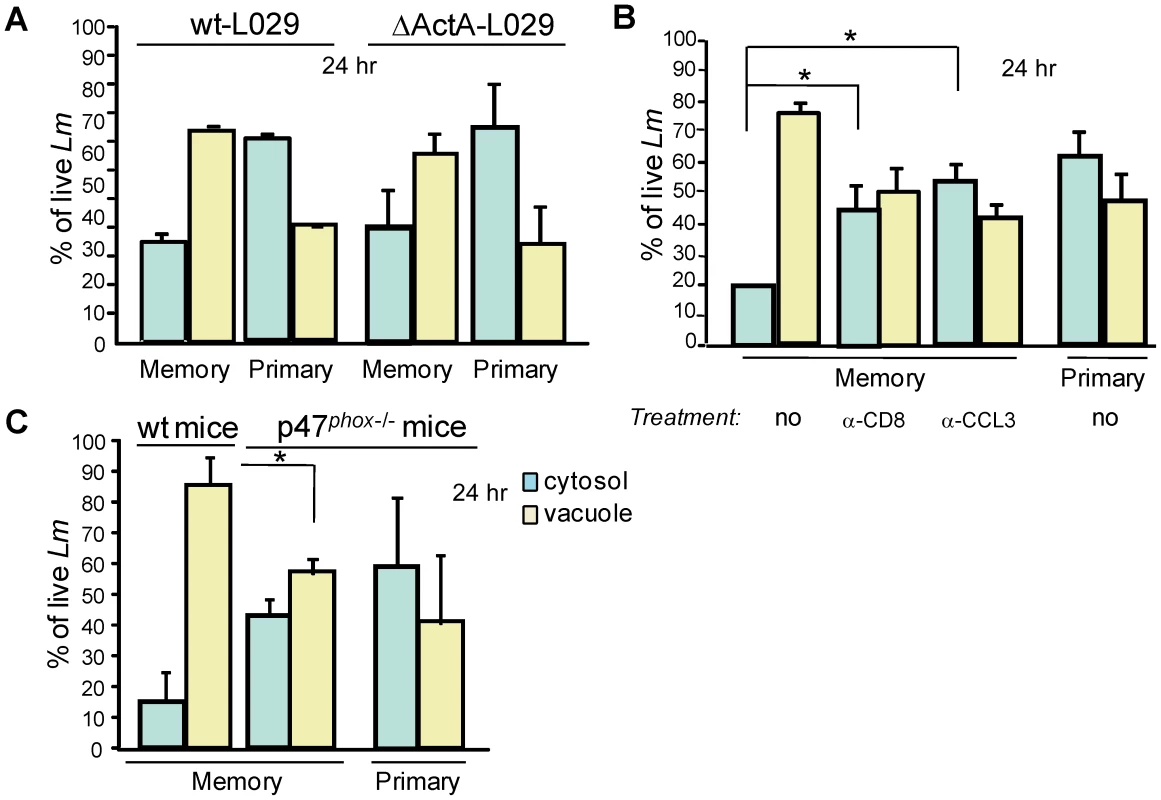
In (A), primary and immune mice (4–6/group) were challenged with 3×10⁵ wt- or ΔActA- L029 Lm and spleens were harvested 24 hrs later and treated as described in figure 5. Histograms show the frequencies (mean +/− SE) of cytosolic (blue bars) and vacuolar (yellow bars) Lm CFUs in phagocytes in a pool of 2 experiments. In (B), primary and memory mice (9–12/group) were treated or not with anti-CD8 or anti-CCL3 before challenge with 3×10⁵ wt-L029 Lm. Histograms indicate as in (A) in a pool of 3 experiments (n = 9). In (C), primary and memory p47phox−/− and C57BL/6 wt control mice (5–6/group) were challenged with 3×10⁵ wt-L029 Lm. Data indicate the number (mean +/− SE) of cytosolic and vacuolar Lm CFUs in phagocytes (n = 5). To demonstrate that Lm relocalization from the cytosol to vacuolar structures indeed depended on CCL3+ memory CD8+ T cells, primary and memory mice were treated with an anti-CD8 depleting mAb, an anti-CCL3 neutralizing or control serum (not shown), and further challenged for 24 hrs with wt-L029 Lm (Figure 6B). CD8+ T cell depletion and CCL3 neutralization both prevented Lm engulfment into such vacuoles of phagocytes from memory mice that exhibited relative frequencies of cytosolic and vacuolar viable Lm similar to that of primary infected animals. Therefore, the subcellular localization of Lm was strongly modified during a memory response and this depended on memory CD8+ T cells and CCL3. This process also correlated with the more efficient bacterial killing occurring in memory mice compared to primary infected animals, which showed 1,920 and 415,200 bacterial CFUs respectively in the spleen 24 hrs after the infection (Figure 4). Overall, these data suggest that Lm destruction in the later phase of infection involves killing mechanisms distinct from those expressed in the primary vacuole of phagocytosis.
We next investigated how such de novo phagosomes that are likely key contributors to Lm destruction during recall infections were formed. Since ROS were required to clear Lm during a memory response (Figure 1B & [6]), we hypothesized that the production of ROS was also involved in Lm re-engulfment to these vacuolar structures of phagocytes from memory mice. For this, wt and p47phox-/- primary or memory mice were challenged with wt-L029 Lm and 24 hrs later the distribution of viable vacuolar and cytosolic bacteria was analyzed (Figure 6C). p47 phox-/- memory mice exhibited similar frequencies of viable Lm in the vacuoles and the cytosol of phagocytes compared to primary infected p47 phox-/- and wt control animals. Therefore, our results show that phagocytes from memory mice receive signals that license them to become more efficient to clear Lm by distinct mechanisms, amongst which is the engulfment of Lm into de novo formed phagosomal structures. This mechanism requires the generation of an oxidative burst promoted via the release of CCL3 by memory CD8+ T cells. Of note, we did not find any substantial differences in the frequencies of phagocytes undergoing cell death (Annexin V+, propidium iodide+) between primary and memory infected mice (Figure S6), likely ruling out increased phagocyte death in memory mice as a mechanism for controlling bacterial growth.
Activation of autophagy in inflammatory monocytes and neutrophils by memory CD8+ T cells
Lm has been shown to be a target for autophagy, a process that can be promoted by the generation of ROS [20], [40]. Since we observed that Lm is engulfed in vacuolar structures of infected phagocytes in a CCL3/ROS-dependent manner, we hypothesized that the phagocytes from memory mice were also primed to kill Lm by autophagy. We therefore monitored the conversion of the well-characterized autophagy marker LC3 inside the phagocytes. During this cellular process, cytosolic-free LC3-I is converted to lipidated LC3-I (LC3-II) that is covalently linked to phospholipids and associated with autophagosomes. This modification alters LC3-I gel migration properties which can be visualized by electrophoresis and western blotting. We assessed the ratio of LC3-II and LC3-I inside inflammatory monocytes, neutrophils and population of macrophages that extracted from the spleens of naïve, primary and memory mice infected for 20 hrs (Figure 7A). In contrast to these macrophages which demonstrated intrinsically high levels of autophagy in naïve uninfected mice (bottom blot), LC3-II/LC3-I ratios in inflammatory monocytes and neutrophils purified from the spleen of memory mice (white bars) were 2 and 1.3 times increased as compared to those from primary infected mice (black bars). This increase in LC3-II/LC3-I ratios most likely reflected the direct consequence of LC3-I lipidation and site specific proteolysis, e.g., conversion of LC3-I into LC3-II that localizes at the membranes of autophagosomes. Of note, we did not observe any significant increase in levels of LC3-I (Figure S7), suggesting that its rate of production was not affected by infection. Using electron microscopy, we looked for the presence of autophagosomes in phagocytes purified by cell sorting from memory mice infected with Lm expressing GFP for 20 hrs [22]. Figure 7B shows representative pictures of infected phagocytes containing Lm engulfed inside vacuolar structures that exhibit the characteristic double membrane feature of autophagosomes.
Fig. 7. The reactive oxygen species generated inside inflammatory monocytes and neutrophils from memory mice induce autophagy. 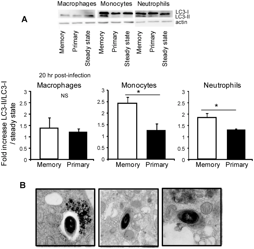
(A) Primary (black bars) and memory mice (white bars) (25/group) were challenged or not with 3×10⁵ wt-L029 Lm. 20 hrs after the infection, spleen cells (from 5 mice/group) were pooled, and flow-sorted inflammatory monocytes, neutrophils and macrophages lysed and each lysate separated on 15% SDS–PAGE and subsequently analyzed with anti-LC3 and anti-actin mAbs (control). (A) Data show representative images (upper) and the quantification histograms (lower) depicting the fold increase of LC3-II/LC3-I ratios (mean +/− SE) measured for each condition and cell subsets compared to the steady state in a pool of 2 to 5 independent experiments. In (B) primary and memory mice (7/group) were challenged with 5×10⁵ wt Lm expressing GFP. 20 hrs later, spleen cells (5 mice/group) were pooled and flow-sorted CD11b+GFP+ infected cells were fixed with glutaraldehyde, and analyzed by electron microscopy. Representative EM images of autophagosomes formation exhibiting double membranes around the bacteria are shown. To formally link the induction of autophagy in monocytes and neutrophils from memory mice with the generation of an oxidative burst and the presence of CCL3+ memory CD8+ T cells, wt control or p47phox−/− primary and memory mice were treated with an anti-CD8 mAb or an anti-CCL3 serum and challenged with wt Lm (Figure S8 and not shown). CD8+ T cell depletion, neutralization of CCL3 or p47phox deficiency all prevented the induction of autophagy in inflammatory monocytes and neutrophils as compared to relevant controls. Collectively, our results show that innate inflammatory monocytes and neutrophils were able, with the help of the CCL3+ memory CD8+ T cells, to specifically induce efficient autophagosome formation during a memory response, and this allows the rapid control of the growth and the clearance of intracellular Lm.
Discussion
Our study highlights that several effector functions of phagocytes are strongly modulated by memory CD8+ T cells in the course of a recall infection in vivo and that this leads to most efficient pathogen destruction. We show that bacterial killing involves activation of higher numbers of monocytes and neutrophils, which produce higher levels of ROS on a per cell basis during the secondary compared to the primary infection. ROS are likely inducing the rapid increase of the pH of the primary vacuoles of phagocytosis, as well as augmented levels of cellular autophagy, both allowing for Lm clearance. Collectively our results show that the cells from the innate immune system behave differently in the course of a recall infection by integrating signals from memory CD8+ T cells which stimulate them to express optimized antimicrobial effector functions. Recent studies also documented enhanced effector/memory CD4+ T cell-mediated clearance of pathogens by innate immune cells [8]–[9]; it is therefore conceivable that comparable antimicrobial protective mechanisms inside effector phagocytes are involved. Along similar lines, an elegant study documented that during lymphochoriomeningitis-induced meningitis, effector CD8+ T cells can promote the recruitment of pathogenic monocytes and neutrophils, leading to accelerated fatal outcome [41].
The current dogma is that only classical T and B lymphocytes that belong to the adaptive immune system are able to differentiate into long-lived memory cells exhibiting qualitatively improved functional properties. In fact, immunological memory is reflected by a stronger proliferation and expression of several effector functions, such as the secretion of specific cytokines and chemokines and the cytolysis of infected cells. In contrast, effector cells of the innate immune system like monocytes, macrophages and neutrophils are believed to mediate a rapid and antigen non-specific immune response that is qualitatively and quantitatively comparable each time the same pathogen is encountered. Macrophages have nevertheless been shown to undergo an adaptive-like response by acquiring distinct patterns of TLR-induced chromatin modifications that include modifications associated with the priming of antimicrobial effectors [42]. In the present work, we found that production of a chemokine by memory CD8+ T cells induced a strikingly distinct response of inflammatory monocytes and neutrophils. Indeed, we observed higher frequencies of ROS-producing inflammatory monocytes and neutrophils, an improved capacity to produce ROS and the upregulation of autophagy, a process also involved in intracellular pathogen clearance. Therefore, our data and that from others [7], [9], [41] promote the concept that the innate immune response during a secondary antigenic encounter can be regulated, either in a cell autonomous manner [42] or in response to lymphocyte-derived cues.
Interestingly, the licensing phenomenon we describe here seems exclusively observed for inflammatory monocytes and neutrophils and likely not for the population of macrophages extracted from the spleen. One explanation could be that the primary function of monocytes and neutrophils is to circulate via the blood and the lymphatic system and patrol the body to permanently sense the tissue environment in search for danger signals [29], [43]. Together, these cell types represent the vast majority of the blood-derived leucocytes and are rapidly attracted to damaged or infected tissues. Another possibility is that splenic monocytes, that may represent an important reservoir of resident monocytes possibly distinct from the blood monocytes, have intrinsically different functional properties [44]. Monocytes indeed exhibit a very dynamic plasticity and are able to differentiate into several functionally distinct subsets of effector cells within inflamed tissues [45]. Therefore, it is conceivable that these cells are more sensitive to cytokine/chemokine-mediated modulation of their fate in vivo compared to tissue-resident macrophages, which are terminally differentiated cells usually involved in steady-state tissue homeostasis.
What mechanisms are responsible for the improved immune response of innate phagocytes during secondary Lm infection? We recently found that during the recall infection, memory CD8+ T cells form transient “effector clusters” with inflammatory monocytes and neutrophils in the spleen [46]. These observations provide a spatio-temporal explanation to the subsequent cascade of effector functions that ultimately result in an efficient control of Lm burden during recall infection. In such clusters, memory CD8+ T cell-derived cytokines are concentrated, which likely favors the rapid and efficient integration of inflammatory signals by the phagocytes.
At the molecular level, it will be interesting to investigate whether the augmented production of ROS observed in inflammatory monocytes and neutrophils results from increased phosphorylation of the cytosolic regulatory protein p47phox leading to NADPH oxidase activation [47]. The improved response of phagocytes could then be attributed to post-translational regulation mechanisms. ROS, and more specifically O2−, can act as signaling molecules to induce cellular autophagy [40], a mechanism that is increasingly described as an important antimicrobial effector mechanism to kill pathogens. The increased level of ROS produced in the course of a memory response can also induce higher pH inside the vacuoles of inflammatory monocytes and neutrophils, and better bacterial killing. Higher pH can induce ion fluxes across the vacuolar membrane, which displaces strongly bound enzymes from the negatively charged proteoglycan granule matrix and allows them to digest and kill the microorganisms [14].
Collectively our data illustrate that the innate immune response and several antimicrobial effector functions of phagocyte populations can be regulated in the course of a memory response. ROS generation induced upon CCL3 secretion by reactivated memory CD8+ T cells likely provides the major signaling molecules promoting the expression of antimicrobial activities of both inflammatory monocytes and neutrophils. As we previously found, such processes also mediate the bystander killing of an unrelated intracellular pathogen [6]. Since we and others have shown that the modulation of the functions expressed by these phagocytes is potentially important for immunotherapeutic and vaccination strategies, it is critical to achieve a better knowledge on how such innate effector cells function in the context of distinct infections.
Materials and Methods
Ethics statement
This study was carried out in strict accordance with the recommendations in the Guide for the Care and Use of Laboratory Animals of the Committee of Animal Care and Use of the Regional Cote d'Azur. The protocol was approved by the Committee on the Ethics of Animal Experiments of the Institut de Pharmacologie Moléculaire et Cellulaire where the study was performed (Permit Number: B-06-152-5, delivered by the Veterinary Services of the Alpes-Maritimes Prefecture) and by the animal use committees at the Albert Einstein College of Medicine. All efforts were made to minimize suffering and provide humane treatment to the animals included in the study.
Mice
Wt BALB/cByJ (Charles River Labs) and C57BL/6J (Jackson labs) 6–8 wk-old female mice, were used in all experiments unless otherwise indicated. p47phox−/− mice were obtained from European Mouse Mutant Archive (EMMA) and CX3CR1+/− mice [48] on the C57BL/6 background were obtained from F. Geissmann's laboratory (King's College London, London). iNOS−/− mice were purchased from CDTA (CNRS, Orléans). Transgenic mice were housed and bred in our SPF animal facility.
Bacteria
The Listeria monocytogenes (Lm) 10403 s wt strain was used in all experiments. The wt-GFP, ΔLLO, wt-L029 and ΔActA Lm are on the 10403 s background and have been previously described [22], [39]. The ΔActA-L029 was generated by transfection of the pLCR plasmid that expresses the chloramphenicol resistance gene under the ActA promoter. The wt-L029 Lm and the pLCR plasmid are a kind gift from Dan Portnoy (UCSB, CA, USA). The wt strains exhibit a LD50 of 3×10⁴ in BALB/c mice. HKLM was prepared as previously described [49]. All Lm were prepared from clones grown from organs of infected mice and kept at −80°C.
Infection of mice and measure of protective immunity and mice survival
For Lm infections, bacteria were grown to a logarithmic phase (OD600 = 0.05–0.15) in Broth Heart Infusion medium (Sigma), diluted in PBS and injected i.v. into lateral tail vein. In all experiments, mice were primary immunized with a 0.1xLD50 of bacteria (3×10³). Secondary infections were carried out one month later with indicated bacteria. To measure Lm titers in spleen and liver, organs were dissociated in 0.1% X-100 Triton (Sigma) and serial dilutions plated onto BHI media plates. To determine the number and frequency of viable Lm localized inside the vacuole or the cytosol, spleens were dissociated in RPMI1640 (Gibco) 5% FCS and each cell suspension was split in two and either incubated or not with 10 µg/ml of chloramphenicol at 37°C for 45 min. After centrifugation, cell pellets were resuspended in 0.1% triton X-100 and serial dilutions plated onto BHI media plates. The next day, we determined the total number (vacuolar and cytosolic) of viable bacteria by counting the colony forming units (CFU) from untreated samples. The number of cytosolic bacteria was determined by counting the CFU from chloramphenicol-treated cells. By subtracting these numbers, we obtained the number of vacuolar bacteria. Lm CFUs in these cells were determined as described above.
In vivo neutralization and depletion experiments
For neutralization of CCL3, mice were treated with 75 µg of anti-CCL3 or goat IgG control i.v at the time of the secondary infection and 24 h later. For neutralization of IFN-γ, mice received one injection of 500 µg of the mAb XMG1.2 concomitantly to the recall infection. For depletion of CD8+ T cells, mice were injected daily 3 times with 100 µg of the anti-CD8β H-35 mAb or its control isotype i.p and further infected one day after.
Antibodies and reagents
Anti-F4/80 (A3-1)-fluorescein isothiocyanate (FITC) was purchased from Caltag Laboratories. The following mAbs were purchased from BD Pharmingen:, anti-CD11b (M1/70) -phycoerythrin (PE), -peridinin chlorophyll protein (PcP) or -allophycocyanin (APC), anti-TCR-PE (H57-597), anti-NK1.1-PE (PK136), anti-CD19-PE (MB19-1), anti-Ly-6G-PE (1A8), anti-Ly-6C-FITC and biotine (AL-21), anti-CD11c-PE and APC (HL3), anti-TNF-α-APC (MP6-XT22) and control rat IgG1 mAb, AnnexinV and propidium iodide. Anti-NOS-2 (M-19) polyclonal rabbit Abs were purchased from Santa Cruz Biotechnology. Goat anti-rabbit-Alexa647 was from Molecular Probes.
Cell suspensions
Organs were cut in small pieces and incubated at 37°C for 20 min in HBSS medium (Gibco) containing 4000 U/ml of collagenase I (Gibco) and 0.1 mg/ml of DNase I (Roche). Red blood cells were lysed for 2–3 min in 170 mM NH4Cl, 17 mM Tris HCl pH 7.4.
Antibody staining and flow cytometry
Cells were stained with the specified Abs in PBS 0.5% of BSA (FACS buffer). For intracellular TNF-α staining, splenocytes were incubated at 37°C 5%CO2 for 3–4 hrs in RPMI1640 (Gibco) 5% FCS, 2 µg/ml Golgi Plug (BD Pharmingen) with or without 5×10⁸ HKLM/ml. Cells were incubated for 20 min on ice with the indicated cell surface marker mAbs, fixed in 1% PFA FACS buffer for 20 min on ice, permeabilized for 30 min in 1XPerm/Wash (BD Pharmingen). For intracellular staining of TNF-α cells were incubated for 20 min on ice in FACS buffer containing anti-TNF-α or control rat IgG1. For intracellular staining of iNOS, cells were incubated for 20 min on ice in FACS buffer containing anti-iNOS rabbit polyclonal, or control normal goat IgG and staining was revealed using goat anti-rabbit Alexa647 mAb. In all cases, cells were washed, fixed for 30 minutes in 1% PFA FACS buffer and analyzed on a FACScalibur cytofluorometer (Becton Dickinson, BD). When indicated, cells were sorted on a FACSvantage SE cell sorter (Becton Dickinson).
Assay for the production of ROS
5−10×106 splenocytes were incubated for 3 h at 37°C and 5%CO2 in RPMI1640 containing 5% FCS with 5×10⁸ HKLM/ml and 160 µM hydroethidine (Polysciences). Hydroethidine is oxidized by ROS in red fluorescent ethidium bromide (EB) therefore allowing for the detection of ROS-producing cells. Cells were washed in FACS buffer and stained for expression of cell surface markers.
Measure of phagosomal pH
This protocol was adapted from Savina et al. [34]–[35]. 50 to 70 µl of 3 micrometers of polybeads amino (Molecular probes) were covalently coupled with 50 µl of 50 mg/ml FITC (pH sensitive) (Fluorescein isothio isomer 1, Sigma F7250) and 50 µl of 1 mg/ml FluoProbes 647 (pH insensitive) (Fluo Probes, Molecular Probes FP-AK7740) in 400 µl of sodium hydrogen carbonate buffer at pH 8.0 for 2 h at room temperature. After extensively washing with glycine 100 mM, the beads were suspended in PBS. Cells were then pulsed with the coupled beads for 20 min, extensively washed in cold PBS and stained on ice for CD11b, Ly-6C or F4/40 surface markers and immediately analyzed by FACS, by selectively gating on the cells that have phagocytosed one latex bead. The ratio of the mean fluorescence intensity (MFI) emission between the two dyes was determined. Values were compared with a standard curve obtained by resuspending the cells that had phagocytosed beads for 20 min at a fixed pH (ranging from pH 5.5 to 8.0) and containing 1XPerm/Wash (BD Pharmingen). The cells were immediately analyzed by FACS to determine the emission ratio of the two fluorescent probes for each pH value.
Western blot analysis
PBS-injected or wt immunized BALB/c, C57BL/6 wt or p47phox−/− mice were sacrificed and spleen cells were positively enriched using anti-CD11b-specific MACS beads (Myltenil) according to the standard manufacturer protocol and further flow-cell sorted on expression of CD11b, F4/80 and Ly-6G cell surface markers. Inflammatory monocytes were defined as CD11bmed/highLy-6G-F4/80med cells, neutrophils as CD11bhighLy-6GhighF4/80- cells and macrophages as CD11bmedLy-6GmedF4/80high cells (purity>87%). The same amount of flow-sorted cells (2−4×10⁶) were washed in PBS and pellets were resuspended in lysis buffer pH 8.0 (Tris-HCl 50 mM, NaCl (200 mM), EDTA 5 mM, 0.5% Triton X-100, 0.5% deoxycholic acid) for 45 min at 4°C. Homogenates were centrifuged for 15 min at 20,000xg and lysate supernatants were analyzed by SDS-PAGE, transferred to PVDF membranes, blocked and probed with a rabbit anti-LC3 (2 µg/ml) (Novus Biologicals), a rabbit anti-actin (0.5 µg/ml) (Sigma), a anti-rabbit peroxidase (1∶10,000) (Beckman Coulter). Western blot quantification was performed with the ImageJ software.
Electron microscopy
Flow-sorted CD11b+GFP+ infected cells were fixed with 1.6% glutaraldehyde in 100 mM phosphate buffer pH 7.5. Cells were incubated at 4°C in 100 mM cacodylate buffer pH 7.0 containing 1% osmium tetroxyde. Cellular pellets were washed with distilled water and incubated for 2 hrs with 0.5% uranyl acetate buffer at room temperature in the dark. Cells were washed in water; pellets were dehydrated in increasing acetone series and embedded in epoxy resins. Blocks were thin-sectioned using standard procedures and contrasted for the observation on a Philips CM12 electron microscope.
Statistics
Statistical significance was calculated using an unpaired Mann-Whitney test and Instat software, except if specified differently in the legend of the figure. All P values of 0.05 or less were considered significant and are referred to as such in the text.
Supporting Information
Zdroje
1. HartyJTTvinnereimARWhiteDW 2000 CD8+ T cell effector mechanisms in resistance to infection. Annu Rev Immunol 18 275 308
2. MasopustDVezysVMarzoALLefrancoisL 2001 Preferential localization of effector memory cells in nonlymphoid tissue. Science 291 2413 2417
3. GuidottiLGChisariFV 2001 Noncytolytic control of viral infections by the innate and adaptive immune response. Annu Rev Immunol 19 65 91
4. CowleySCSedgwickJDElkinsKL 2007 Differential requirements by CD4+ and CD8+ T cells for soluble and membrane TNF in control of Francisella tularensis live vaccine strain intramacrophage growth. J Immunol 179 7709 7719
5. MullerIKropfPEtgesRJLouisJA 1993 Gamma interferon response in secondary Leishmania major infection: role of CD8+ T cells. Infect Immun 61 3730 3738
6. Narni-MancinelliECampisiLBassandDCazarethJGounonP 2007 Memory CD8+ T cells mediate antibacterial immunity via CCL3 activation of TNF/ROI+ phagocytes. J Exp Med 204 2075 2087
7. BartonESWhiteDWCathelynJSBrett-McClellanKAEngleM 2007 Herpesvirus latency confers symbiotic protection from bacterial infection. Nature 447 326 329
8. StruttTMMcKinstryKKDibbleJPWinchellCKuangY 2010 Memory CD4+ T cells induce innate responses independently of pathogen. Nat Med 16 558 564 551p following 564
9. ZhangZClarkeTBWeiserJN 2009 Cellular effectors mediating Th17-dependent clearance of pneumococcal colonization in mice. J Clin Invest 119 1899 1909
10. GeissmannFAuffrayCPalframanRWirrigCCioccaA 2008 Blood monocytes: distinct subsets, how they relate to dendritic cells, and their possible roles in the regulation of T-cell responses. Immunol Cell Biol 86 398 408
11. SerbinaNVJiaTHohlTMPamerEG 2008 Monocyte-mediated defense against microbial pathogens. Annu Rev Immunol 26 421 452
12. IshiiKJKoyamaSNakagawaACobanCAkiraS 2008 Host innate immune receptors and beyond: making sense of microbial infections. Cell Host Microbe 3 352 363
13. HenryRMHoppeADJoshiNSwansonJA 2004 The uniformity of phagosome maturation in macrophages. J Cell Biol 164 185 194
14. SegalAW 2005 How neutrophils kill microbes. Annu Rev Immunol 23 197 223
15. BabiorBM 2000 Phagocytes and oxidative stress. Am J Med 109 33 44
16. NathanCDingA 2010 SnapShot: Reactive Oxygen Intermediates (ROI). Cell 140 951 951 e952
17. MacMickingJXieQWNathanC 1997 Nitric oxide and macrophage function. Annu Rev Immunol 15 323 350
18. IsmailNOlanoJPFengHMWalkerDH 2002 Current status of immune mechanisms of killing of intracellular microorganisms. FEMS Microbiol Lett 207 111 120
19. KirkegaardKTaylorMPJacksonWT 2004 Cellular autophagy: surrender, avoidance and subversion by microorganisms. Nat Rev Microbiol 2 301 314
20. PyBFLipinskiMMYuanJ 2007 Autophagy limits Listeria monocytogenes intracellular growth in the early phase of primary infection. Autophagy 3 117 125
21. WalkerDHPopovVLCrocquet-ValdesPAWelshCJFengHM 1997 Cytokine-induced, nitric oxide-dependent, intracellular antirickettsial activity of mouse endothelial cells. Lab Invest 76 129 138
22. MurailleENarni-MancinelliEGounonPBassandDGlaichenhausN 2007 Cytosolic expression of SecA2 is a prerequisite for long-term protective immunity. Cell Microbiol 9 1445 1454
23. IdoyagaJSudaNSudaKParkCGSteinmanRM 2009 Antibody to Langerin/CD207 localizes large numbers of CD8alpha+ dendritic cells to the marginal zone of mouse spleen. Proc Natl Acad Sci U S A 106 1524 1529
24. CookDNSmithiesOStrieterRMFrelingerJASerodyJS 1999 CD8+ T cells are a biologically relevant source of macrophage inflammatory protein-1 alpha in vivo. J Immunol 162 5423 5428
25. KaoCOestreichKJPaleyMACrawfordAAngelosantoJM 2011 Transcription factor T-bet represses expression of the inhibitory receptor PD-1 and sustains virus-specific CD8+ T cell responses during chronic infection. Nat Immunol 12 663 671
26. WirthTCXueHHRaiDSabelJTBairT 2010 Repetitive antigen stimulation induces stepwise transcriptome diversification but preserves a core signature of memory CD8(+) T cell differentiation. Immunity 33 128 140
27. WhiteDWMacNeilABuschDHPilipIMPamerEG 1999 Perforin-deficient CD8+ T cells: in vivo priming and antigen-specific immunity against Listeria monocytogenes. J Immunol 162 980 988
28. MessinghamKABadovinacVPJabbariAHartyJT 2007 A role for IFN-gamma from antigen-specific CD8+ T cells in protective immunity to Listeria monocytogenes. J Immunol 179 2457 2466
29. GeissmannFJungSLittmanDR 2003 Blood monocytes consist of two principal subsets with distinct migratory properties. Immunity 19 71 82
30. SerbinaNVSalazar-MatherTPBironCAKuzielWAPamerEG 2003 TNF/iNOS-producing dendritic cells mediate innate immune defense against bacterial infection. Immunity 19 59 70
31. FlannaganRSCosioGGrinsteinS 2009 Antimicrobial mechanisms of phagocytes and bacterial evasion strategies. Nat Rev Microbiol 7 355 366
32. ReevesEPLuHJacobsHLMessinaCGBolsoverS 2002 Killing activity of neutrophils is mediated through activation of proteases by K+ flux. Nature 416 291 297
33. SegalAWGeisowMGarciaRHarperAMillerR 1981 The respiratory burst of phagocytic cells is associated with a rise in vacuolar pH. Nature 290 406 409
34. SavinaAVargasPGuermonprezPLennonAMAmigorenaS 2010 Measuring pH, ROS production, maturation, and degradation in dendritic cell phagosomes using cytofluorometry-based assays. Methods Mol Biol 595 383 402
35. SavinaAJancicCHuguesSGuermonprezPVargasP 2006 NOX2 controls phagosomal pH to regulate antigen processing during crosspresentation by dendritic cells. Cell 126 205 218
36. DramsiSCossartP 2002 Listeriolysin O: a genuine cytolysin optimized for an intracellular parasite. J Cell Biol 156 943 946
37. PortnoyDAAuerbuchVGlomskiIJ 2002 The cell biology of Listeria monocytogenes infection: the intersection of bacterial pathogenesis and cell-mediated immunity. J Cell Biol 158 409 414
38. Vazquez-BolandJAKuhnMBerchePChakrabortyTDominguez-BernalG 2001 Listeria pathogenesis and molecular virulence determinants. Clin Microbiol Rev 14 584 640
39. MoorsMALevittBYoungmanPPortnoyDA 1999 Expression of listeriolysin O and ActA by intracellular and extracellular Listeria monocytogenes. Infect Immun 67 131 139
40. Scherz-ShouvalRShvetsEFassEShorerHGilL 2007 Reactive oxygen species are essential for autophagy and specifically regulate the activity of Atg4. Embo J 26 1749 1760
41. KimJVKangSSDustinMLMcGavernDB 2009 Myelomonocytic cell recruitment causes fatal CNS vascular injury during acute viral meningitis. Nature 457 191 195
42. FosterSLHargreavesDCMedzhitovR 2007 Gene-specific control of inflammation by TLR-induced chromatin modifications. Nature 447 972 978
43. AuffrayCFoggDGarfaMElainGJoin-LambertO 2007 Monitoring of blood vessels and tissues by a population of monocytes with patrolling behavior. Science 317 666 670
44. SwirskiFKNahrendorfMEtzrodtMWildgruberMCortez-RetamozoV 2009 Identification of splenic reservoir monocytes and their deployment to inflammatory sites. Science 325 612 616
45. GeissmannFManzMGJungSSiewekeMHMeradM 2010 Development of monocytes, macrophages, and dendritic cells. Science 327 656 661
46. BajenoffMNarni-MancinelliEBrauFLauvauG 2010 Visualizing early splenic memory CD8+ T cells reactivation against intracellular bacteria. PLoS One 5 e11524
47. DangPMStensballeABoussettaTRaadHDewasC 2006 A specific p47phox -serine phosphorylated by convergent MAPKs mediates neutrophil NADPH oxidase priming at inflammatory sites. J Clin Invest 116 2033 2043
48. JungSAlibertiJGraemmelPSunshineMJKreutzbergGW 2000 Analysis of fractalkine receptor CX(3)CR1 function by targeted deletion and green fluorescent protein reporter gene insertion. Mol Cell Biol 20 4106 4114
49. LauvauGVijhSKongPHorngTKerksiekK 2001 Priming of memory but not effector CD8 T cells by a killed bacterial vaccine. Science 294 1735 1739
Štítky
Hygiena a epidemiológia Infekčné lekárstvo Laboratórium
Článek Genesis of Mammalian Prions: From Non-infectious Amyloid Fibrils to a Transmissible Prion DiseaseČlánek Role of Permissive Neuraminidase Mutations in Influenza A/Brisbane/59/2007-like (H1N1) VirusesČlánek Allelic Variation on Murine Chromosome 11 Modifies Host Inflammatory Responses and Resistance toČlánek Multifaceted Regulation of Translational Readthrough by RNA Replication Elements in a TombusvirusČlánek Latent KSHV Infection of Endothelial Cells Induces Integrin Beta3 to Activate Angiogenic PhenotypesČlánek Controlling Viral Immuno-Inflammatory Lesions by Modulating Aryl Hydrocarbon Receptor Signaling
Článok vyšiel v časopisePLOS Pathogens
Najčítanejšie tento týždeň
2011 Číslo 12- Parazitičtí červi v terapii Crohnovy choroby a dalších zánětlivých autoimunitních onemocnění
- Očkování proti virové hemoragické horečce Ebola experimentální vakcínou rVSVDG-ZEBOV-GP
- Koronavirus hýbe světem: Víte jak se chránit a jak postupovat v případě podezření?
-
Všetky články tohto čísla
- Inhibition of Apoptosis and NF-κB Activation by Vaccinia Protein N1 Occur via Distinct Binding Surfaces and Make Different Contributions to Virulence
- Genesis of Mammalian Prions: From Non-infectious Amyloid Fibrils to a Transmissible Prion Disease
- Kaposi's Sarcoma Herpesvirus microRNAs Target Caspase 3 and Regulate Apoptosis
- Nutritional Immunology: A Multi-Dimensional Approach
- Role of Permissive Neuraminidase Mutations in Influenza A/Brisbane/59/2007-like (H1N1) Viruses
- Vaccinomics and Personalized Vaccinology: Is Science Leading Us Toward a New Path of Directed Vaccine Development and Discovery?
- Symbiont Infections Induce Strong Cytoplasmic Incompatibility in the Tsetse Fly
- Allelic Variation on Murine Chromosome 11 Modifies Host Inflammatory Responses and Resistance to
- Computational and Biochemical Analysis of the Effector AvrBs2 and Its Role in the Modulation of Type Three Effector Delivery
- Granzyme B Inhibits Vaccinia Virus Production through Proteolytic Cleavage of Eukaryotic Initiation Factor 4 Gamma 3
- Association of Activating KIR Copy Number Variation of NK Cells with Containment of SIV Replication in Rhesus Monkeys
- Fungal Virulence and Development Is Regulated by Alternative Pre-mRNA 3′End Processing in
- versus the Host: Remodeling of the Bacterial Outer Membrane Is Required for Survival in the Gastric Mucosa
- Follicular Dendritic Cell-Specific Prion Protein (PrP) Expression Alone Is Sufficient to Sustain Prion Infection in the Spleen
- Autophagy Protein Atg3 is Essential for Maintaining Mitochondrial Integrity and for Normal Intracellular Development of Tachyzoites
- Longevity and Composition of Cellular Immune Responses Following Experimental Malaria Infection in Humans
- Sequential Adaptive Mutations Enhance Efficient Vector Switching by Chikungunya Virus and Its Epidemic Emergence
- Acquisition of Pneumococci Specific Effector and Regulatory Cd4 T Cells Localising within Human Upper Respiratory-Tract Mucosal Lymphoid Tissue
- The Meaning of Death: Evolution and Ecology of Apoptosis in Protozoan Parasites
- Deficiency of a Niemann-Pick, Type C1-related Protein in Is Associated with Multiple Lipidoses and Increased Pathogenicity
- Feeding Cells Induced by Phytoparasitic Nematodes Require γ-Tubulin Ring Complex for Microtubule Reorganization
- Eight RGS and RGS-like Proteins Orchestrate Growth, Differentiation, and Pathogenicity of
- Prion Uptake in the Gut: Identification of the First Uptake and Replication Sites
- Nef Decreases HIV-1 Sensitivity to Neutralizing Antibodies that Target the Membrane-proximal External Region of TMgp41
- Multifaceted Regulation of Translational Readthrough by RNA Replication Elements in a Tombusvirus
- A Temporal Role Of Type I Interferon Signaling in CD8 T Cell Maturation during Acute West Nile Virus Infection
- The Membrane Fusion Step of Vaccinia Virus Entry Is Cooperatively Mediated by Multiple Viral Proteins and Host Cell Components
- HIV-1 Capsid-Cyclophilin Interactions Determine Nuclear Import Pathway, Integration Targeting and Replication Efficiency
- Neonatal CD8 T-cell Hierarchy Is Distinct from Adults and Is Influenced by Intrinsic T cell Properties in Respiratory Syncytial Virus Infected Mice
- Two Novel Transcriptional Regulators Are Essential for Infection-related Morphogenesis and Pathogenicity of the Rice Blast Fungus
- Five Questions about Non-Mevalonate Isoprenoid Biosynthesis
- The Human Cytomegalovirus UL11 Protein Interacts with the Receptor Tyrosine Phosphatase CD45, Resulting in Functional Paralysis of T Cells
- Wall Teichoic Acids of Limit Recognition by the Drosophila Peptidoglycan Recognition Protein-SA to Promote Pathogenicity
- A Novel Role for the NLRC4 Inflammasome in Mucosal Defenses against the Fungal Pathogen
- Inflammasome-dependent Pyroptosis and IL-18 Protect against Lung Infection while IL-1β Is Deleterious
- CNS Recruitment of CD8+ T Lymphocytes Specific for a Peripheral Virus Infection Triggers Neuropathogenesis during Polymicrobial Challenge
- Latent KSHV Infection of Endothelial Cells Induces Integrin Beta3 to Activate Angiogenic Phenotypes
- A Receptor-based Switch that Regulates Anthrax Toxin Pore Formation
- Targeting of Heparin-Binding Hemagglutinin to Mitochondria in Macrophages
- Chikungunya Virus Neutralization Antigens and Direct Cell-to-Cell Transmission Are Revealed by Human Antibody-Escape Mutants
- Ce-Duox1/BLI-3 Generated Reactive Oxygen Species Trigger Protective SKN-1 Activity via p38 MAPK Signaling during Infection in
- Structural Elucidation and Functional Characterization of the Effector Protein ATR13
- Controlling Viral Immuno-Inflammatory Lesions by Modulating Aryl Hydrocarbon Receptor Signaling
- SAMHD1-Deficient CD14+ Cells from Individuals with Aicardi-Goutières Syndrome Are Highly Susceptible to HIV-1 Infection
- Acid Stability of the Hemagglutinin Protein Regulates H5N1 Influenza Virus Pathogenicity
- Cryo Electron Tomography of Herpes Simplex Virus during Axonal Transport and Secondary Envelopment in Primary Neurons
- A Novel Human Cytomegalovirus Locus Modulates Cell Type-Specific Outcomes of Infection
- Juxtamembrane Shedding of AMA1 Is Sequence Independent and Essential, and Helps Evade Invasion-Inhibitory Antibodies
- Pathogenesis and Host Response in Syrian Hamsters following Intranasal Infection with Andes Virus
- IRGM Is a Common Target of RNA Viruses that Subvert the Autophagy Network
- Epstein-Barr Virus Evades CD4 T Cell Responses in Lytic Cycle through BZLF1-mediated Downregulation of CD74 and the Cooperation of vBcl-2
- Quantitative Multicolor Super-Resolution Microscopy Reveals Tetherin HIV-1 Interaction
- Late Repression of NF-κB Activity by Invasive but Not Non-Invasive Meningococcal Isolates Is Required to Display Apoptosis of Epithelial Cells
- Polar Flagellar Biosynthesis and a Regulator of Flagellar Number Influence Spatial Parameters of Cell Division in
- Epstein-Barr Virus Nuclear Antigen 3C Stabilizes Gemin3 to Block p53-mediated Apoptosis
- The Enteropathogenic (EPEC) Tir Effector Inhibits NF-κB Activity by Targeting TNFα Receptor-Associated Factors
- Toward an Integrated Model of Capsule Regulation in
- A Systematic Screen to Discover and Analyze Apicoplast Proteins Identifies a Conserved and Essential Protein Import Factor
- A Host Small GTP-binding Protein ARL8 Plays Crucial Roles in Tobamovirus RNA Replication
- Comparative Pathobiology of Fungal Pathogens of Plants and Animals
- Synergistic Roles of Eukaryotic Translation Elongation Factors 1Bγ and 1A in Stimulation of Tombusvirus Minus-Strand Synthesis
- Engineered Immunity to Infection
- Inflammatory Monocytes and Neutrophils Are Licensed to Kill during Memory Responses
- Sialidases Affect the Host Cell Adherence and Epsilon Toxin-Induced Cytotoxicity of Type D Strain CN3718
- Eurasian-Origin Gene Segments Contribute to the Transmissibility, Aerosol Release, and Morphology of the 2009 Pandemic H1N1 Influenza Virus
- SARS Coronavirus nsp1 Protein Induces Template-Dependent Endonucleolytic Cleavage of mRNAs: Viral mRNAs Are Resistant to nsp1-Induced RNA Cleavage
- Identification and Characterization of a Novel Non-Structural Protein of Bluetongue Virus
- Functional Analysis of the Kinome of the Wheat Scab Fungus
- Norovirus Regulation of the Innate Immune Response and Apoptosis Occurs via the Product of the Alternative Open Reading Frame 4
- PLOS Pathogens
- Archív čísel
- Aktuálne číslo
- Informácie o časopise
Najčítanejšie v tomto čísle- Controlling Viral Immuno-Inflammatory Lesions by Modulating Aryl Hydrocarbon Receptor Signaling
- Fungal Virulence and Development Is Regulated by Alternative Pre-mRNA 3′End Processing in
- Epstein-Barr Virus Nuclear Antigen 3C Stabilizes Gemin3 to Block p53-mediated Apoptosis
- Engineered Immunity to Infection
Prihlásenie#ADS_BOTTOM_SCRIPTS#Zabudnuté hesloZadajte e-mailovú adresu, s ktorou ste vytvárali účet. Budú Vám na ňu zasielané informácie k nastaveniu nového hesla.
- Časopisy


