-
Články
- Časopisy
- Kurzy
- Témy
- Kongresy
- Videa
- Podcasty
The Anti-Sigma Factor TcdC Modulates Hypervirulence in an Epidemic BI/NAP1/027 Clinical Isolate of
Nosocomial infections are increasingly being recognised as a major patient safety issue. The modern hospital environment and associated health care practices have provided a niche for the rapid evolution of microbial pathogens that are well adapted to surviving and proliferating in this setting, after which they can infect susceptible patients. This is clearly the case for bacterial pathogens such as Methicillin Resistant Staphylococcus aureus (MRSA) and Vancomycin Resistant Enterococcus (VRE) species, both of which have acquired resistance to antimicrobial agents as well as enhanced survival and virulence properties that present serious therapeutic dilemmas for treating physicians. It has recently become apparent that the spore-forming bacterium Clostridium difficile also falls within this category. Since 2000, there has been a striking increase in C. difficile nosocomial infections worldwide, predominantly due to the emergence of epidemic or hypervirulent isolates that appear to possess extended antibiotic resistance and virulence properties. Various hypotheses have been proposed for the emergence of these strains, and for their persistence and increased virulence, but supportive experimental data are lacking. Here we describe a genetic approach using isogenic strains to identify a factor linked to the development of hypervirulence in C. difficile. This study provides evidence that a naturally occurring mutation in a negative regulator of toxin production, the anti-sigma factor TcdC, is an important factor in the development of hypervirulence in epidemic C. difficile isolates, presumably because the mutation leads to significantly increased toxin production, a contentious hypothesis until now. These results have important implications for C. difficile pathogenesis and virulence since they suggest that strains carrying a similar mutation have the inherent potential to develop a hypervirulent phenotype.
Published in the journal: The Anti-Sigma Factor TcdC Modulates Hypervirulence in an Epidemic BI/NAP1/027 Clinical Isolate of. PLoS Pathog 7(10): e32767. doi:10.1371/journal.ppat.1002317
Category: Research Article
doi: https://doi.org/10.1371/journal.ppat.1002317Summary
Nosocomial infections are increasingly being recognised as a major patient safety issue. The modern hospital environment and associated health care practices have provided a niche for the rapid evolution of microbial pathogens that are well adapted to surviving and proliferating in this setting, after which they can infect susceptible patients. This is clearly the case for bacterial pathogens such as Methicillin Resistant Staphylococcus aureus (MRSA) and Vancomycin Resistant Enterococcus (VRE) species, both of which have acquired resistance to antimicrobial agents as well as enhanced survival and virulence properties that present serious therapeutic dilemmas for treating physicians. It has recently become apparent that the spore-forming bacterium Clostridium difficile also falls within this category. Since 2000, there has been a striking increase in C. difficile nosocomial infections worldwide, predominantly due to the emergence of epidemic or hypervirulent isolates that appear to possess extended antibiotic resistance and virulence properties. Various hypotheses have been proposed for the emergence of these strains, and for their persistence and increased virulence, but supportive experimental data are lacking. Here we describe a genetic approach using isogenic strains to identify a factor linked to the development of hypervirulence in C. difficile. This study provides evidence that a naturally occurring mutation in a negative regulator of toxin production, the anti-sigma factor TcdC, is an important factor in the development of hypervirulence in epidemic C. difficile isolates, presumably because the mutation leads to significantly increased toxin production, a contentious hypothesis until now. These results have important implications for C. difficile pathogenesis and virulence since they suggest that strains carrying a similar mutation have the inherent potential to develop a hypervirulent phenotype.
Introduction
C. difficile is the causative agent of a spectrum of gastrointestinal diseases, collectively known as C. difficile infections, or CDI, that are induced by treatment with antibiotics that disrupt the normal gastrointestinal microbiota. CDI can range from mild diarrhoea, through moderately serious disease, to severe life-threatening pseudomembranous colitis, a chronic, often fatal, gastrointestinal disease [1]. During the past decade, there has been an astonishing increase in the rate and prevalence of C. difficile infections in many parts of the world, including the UK, USA, Canada and Europe, largely due to the emergence of a “hypervirulent” or epidemic group of isolates belonging to the BI/NAP1/027 category [2], [3]. These strains are highly resistant to fluoroquinolones [3] and are associated with more severe disease and higher mortality rates [4]–[7]. C. difficile now also causes disease in those previously not at risk, such as children and pregnant women, with community-associated C. difficile disease being increasingly common [8]–[10].
The reasons for the emergence of these strains, and for their increased virulence, remain largely speculative. The use of fluoroquinolones, and the emergence of fluoroquinolone resistant strains, are undoubtedly driving factors in these new epidemics [11], however, the reasons for the heightened virulence and persistence of these strains are unknown. Genotypic and phenotypic comparison of the hypervirulent BI/NAP1/027 isolates to historical strains has identified numerous differences that may contribute to hypervirulence. Phenotypically, these differences may include the production of a toxin known as binary toxin, or CDT [3], and a higher sporulation rate [12]. Whole genome comparisons have identified numerous genetic differences with BI/NAP1/027 strains having an additional 234 genes compared to the well characterised strain 630 [13], including five unique genetic regions that are absent from both strain 630 and non-epidemic 027 strains [4]. Fundamentally, however, the factors directly resulting in the development of hypervirulence by these strains remain unknown.
The major virulence factors of C. difficile are two members of the large clostridial cytotoxin family, toxin A and toxin B, encoded by the tcdA and tcdB genes, respectively, which are potent monoglucosyltransferases that irreversibly modify members of the Rho family of host regulatory proteins [14]. Two recent studies definitively showed that toxin B plays a major role in the virulence of C. difficile [15], [16]. The role of toxin A in disease was less clear however, with conflicting data concerning toxin A reported [15], [16].
Epidemic strains are reported to produce significantly more toxin A and toxin B than other strains [2]. The tcdA and tcdB genes are located on the chromosome within a region known as the pathogenicity locus or PaLoc [17]. In addition to tcdA and tcdB, the PaLoc encodes three additional genes designated tcdR, tcdE and tcdC, which encode an alternative sigma factor, TcdR [18], a putative holin, TcdE [19], and an anti-sigma factor, TcdC [20], respectively. The expression of toxins A and B is controlled in a complex manner by several factors, including TcdR and TcdC. TcdC is thought to negatively regulate toxin production by interacting with TcdR or with TcdR-containing RNA polymerase holoenzyme or both [20], TcdR is essential for toxin production [18]. BI/NAP1/027 C. difficile strains have a nonsense mutation in tcdC, which results in the production of a truncated protein that no longer negatively regulates TcdR. This mutation is postulated to be responsible for the increased toxin production observed in vitro in these strains [2]. Accordingly, this observation has prompted debate over the importance of the tcdC mutation in the hypervirulent phenotype. However, there is currently a lack of experimental evidence to support this hypothesis, with inconsistent reports in the published literature [20]–[22].
Despite their important impact worldwide on public health little is known about the virulence factors of BI/NAP1/027 strains and many important questions about the pathogenesis of disease caused by these strains remain to be answered, especially the role played by TcdC. BI/NAP1/027 isolates have proven difficult to genetically manipulate, which has hampered our ability to study these strains at the molecular level. To address these questions, here we use a novel Tn916-based plasmid conjugation system to facilitate the efficient transfer of plasmids into BI/NAP1/027 strains of C. difficile. Using this system, we have demonstrated conclusively the role of TcdC as a negative regulator of toxin production in C. difficile. Furthermore, using the hamster model of infection, we provide evidence to show that the tcdC mutation found in BI/NAP1/027 strains is an important factor in the development of hypervirulence by these strains. This study is the first to use isogenic strains to identify a factor involved in the development of a hypervirulent phenotype in C. difficile, and also represents the first in vivo demonstration of the role of TcdC in the pathogenesis of C. difficile disease.
Results
Complementation of the tcdC mutation in a BI/NAP1/027 epidemic isolate in trans
To determine if mutation of the tcdC gene in C. difficile BI/NAP1/027 isolates leads to the development of a hypervirulent phenotype it was necessary to construct isogenic BI/NAP1/027 strains that only differed in their ability to produce a functional TcdC protein. To construct the isogenic strains required for this analysis, genetic manipulation of BI/NAP1/027 isolates was required. The genetic manipulation of these strains has proved difficult and attempts to transfer plasmids into BI/NAP1/027 strains using published methods, which rely on RP4-mediated conjugation from Escherichia coli [23]–[27], were not successful, even though transfer of plasmids into the genetically amenable strains JIR8094, an erythromycin sensitive derivative of strain 630 [24], and CD37 was readily achieved (Table S1). To overcome this barrier and to facilitate DNA transfer into the strains of interest, we developed a novel plasmid transfer system that exploits the conjugation apparatus encoded by the broad-host range transposon Tn916.
The oriT region of Tn916 (oriTTn916) [28] was cloned into the catP-containing C. difficile shuttle plasmid pMTL9361Cm [29], generating pDLL4. This plasmid was introduced into C. perfringens strain JIR4225, which contains five copies of Tn916 [30] and plate matings were performed between this donor strain and several C. difficile strains, including a BI/NAP1/027 strain, M7404, which is a Canadian epidemic isolate [29]. Transconjugants from these matings were isolated on medium supplemented with thiamphenicol and cefoxitin. The efficiency of plasmid transfer into strain M7404 was 1.2×10²–4×10⁴ transconjugants/ml of plated culture. Analysis of transconjugants using PCR specific for the catP gene together with restriction analysis confirmed that all putative colonies carried pDLL4 (data not shown), verifying successful plasmid transfer into the BI/NAP1/027 strain M7404. Similar plasmid transfer efficiencies were obtained for numerous other C. difficile strains (Table S1), highlighting the utility of this methodology for the genetic manipulation of clinically relevant strains.
To complement the tcdC mutation in a BI/NAP1/027 strain the intact tcdC gene from strain VPI10463, together with 300 bp of its upstream region, was cloned into the shuttle plasmid pDLL4, generating pDLL17. This plasmid was transferred by Tn916-mediated conjugation from C. perfringens strain JIR4225 to C. difficile strain M7404 as before. PCR was subsequently used to confirm the presence of plasmid pDLL17 in representative transconjugants (data not shown).
To determine whether the presence of pDLL17 complemented the TcdC deficiency of M7404, Western immunoblots using TcdC-specific antibodies were performed. Lysates were collected from the wild-type M7404, the pDLL4-carrying vector control strain M7404(VC) and the pDLL17-tcdC+ strain M7404(tcdC+), as well as strains VPI10463 and the PaLoc-deficient strain VPI11186, which served as positive and negative controls, respectively. An additional control strain, M7404(cured), was generated by serially passaging strain M7404(tcdC+) on non-selective growth medium and curing the plasmid from this strain. Loss of the plasmid was confirmed by sensitivity of the strain to thiamphenicol followed by PCR analysis to verify the absence of several plasmid encoded genes (data not shown). As Figure 1A shows, whilst no TcdC could be detected in the lysates of the negative control strain, the wild-type M7404, M7404(VC) and the plasmid-cured strain M7404(cured), a 34-kDa protein that reacted with TcdC-specific antibodies was detected in lysates from the tcdC+-complemented strain M7404(tcdC+). This band was the same size as the immunoreactive TcdC protein produced by the positive control strain VPI10463, confirming that the tcdC mutation in the BI/NAP1/027 epidemic isolate M7404 was efficiently complemented in trans. Since complementation was performed using a multicopy plasmid, we also quantified TcdC production levels from strain M7404(tcdC+) in comparison to strain VPI10463 using a time-course assay. Previous studies involving transcriptional analysis of PaLoc genes during different growth phases showed that tcdC is expressed in early exponential phase but not in stationary phase, whereas the other PaLoc genes show the opposite expression pattern [31]. VPI10463 (Figure 1B) and M7404(tcdC+) (Figure 1C) exhibited similar TcdC expression patterns, with higher levels of TcdC observed in early exponential phase and negligible amounts detected beyond 16 hours, suggesting that the regulatory regions governing tcdC expression have been retained on the tcdC-carrying fragment used to construct pDLL17. In addition to the kinetics of TcdC expression in strain M7404(tcdC+) mirroring that of VPI10463, a similar amount of protein was also detected at each time point with VPI10463 producing 1.3 - to 1.6-fold more protein (Figure 1B) than M7404(tcdC+) (Figure 1C). Therefore, although tcdC complementation was achieved using a multicopy plasmid vector, a physiologically relevant amount of TcdC protein was expressed during the appropriate growth phases in strain M7404(tcdC+).
Fig. 1. Western blot analysis of TcdC production by wild-type and complemented C. difficile strains. 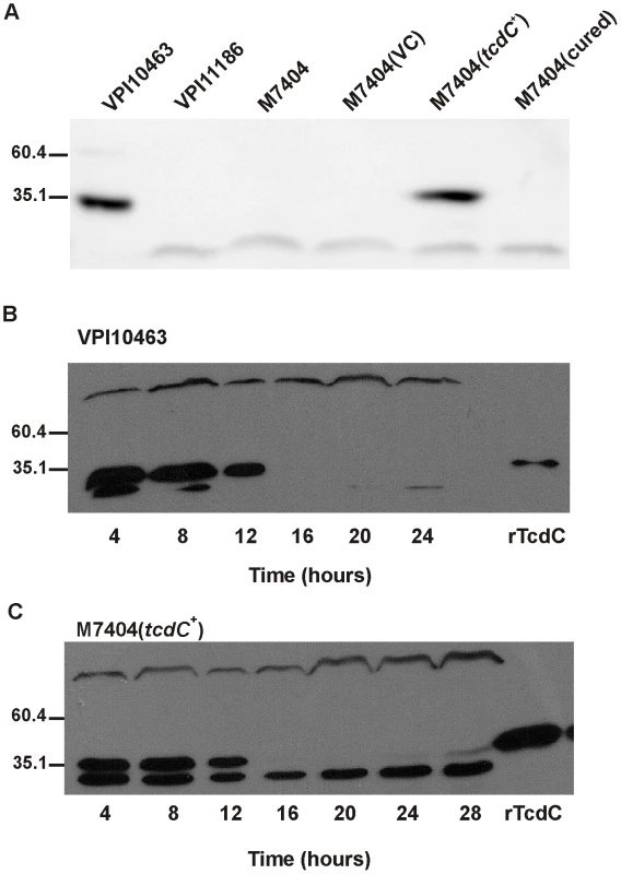
(A) Qualitative analysis of TcdC production. VPI10463 is the positive control strain; VPI11186 is the negative control strain; M7404 is a wild-type Canadian BI/NAP1/027 strain; M7404(VC) is the wild-type strain carrying the shuttle plasmid pDLL4; M7404(tcdC+) is the wild-type strain carrying the tcdC expression plasmid pDLL17 and M7404(cured) is the M7404(tcdC+) strain cured of pDLL17. (B) Time course analysis of TcdC production by the positive control strain C. difficile VPI10463. Samples were taken at the indicated times shown in hours. 60 ng of purified recombinant his-tagged TcdC protein (rTcdC) was used as the positive reference sample. (C) Time course analysis of TcdC production by C. difficile strain M7404(tcdC+). Samples were taken at the indicated times shown in hours. 300 ng of purified recombinant his-tagged TcdC protein (rTcdC) was used as the positive reference sample. Western blots were performed with rabbit TcdC-specific antibodies. Size standards are shown (kDa). TcdC-mediated repression of toxin production in C. difficile
To determine the effect of TcdC on toxin production in strain M7404, a combination of Western immunoblots and cytotoxicity assays were performed using supernatants collected from strain M7404 and isogenic M7404 derivatives carrying the vector pDLL4, pDLL17, the cured strain M7404(cured) and the PaLoc-negative control strain CD37. To assess toxin A production, Western immunoblotting was performed using TcdA-specific antibodies (Figure 2A). The results showed that the presence of the tcdC+ plasmid pDLL17 resulted in a dramatic decrease in the amount of toxin A produced by M7404(tcdC+) when compared to the wild-type strain. By contrast, M7404(cured) produced qualitatively similar levels of toxin A to wild-type, as did M7404 carrying the vector plasmid, whereas the PaLoc-negative strain CD37 produced no detectable toxin A, as expected.
Fig. 2. Analysis of the effect of TcdC complementation on toxin production and PaLoc gene expression by C. difficile. 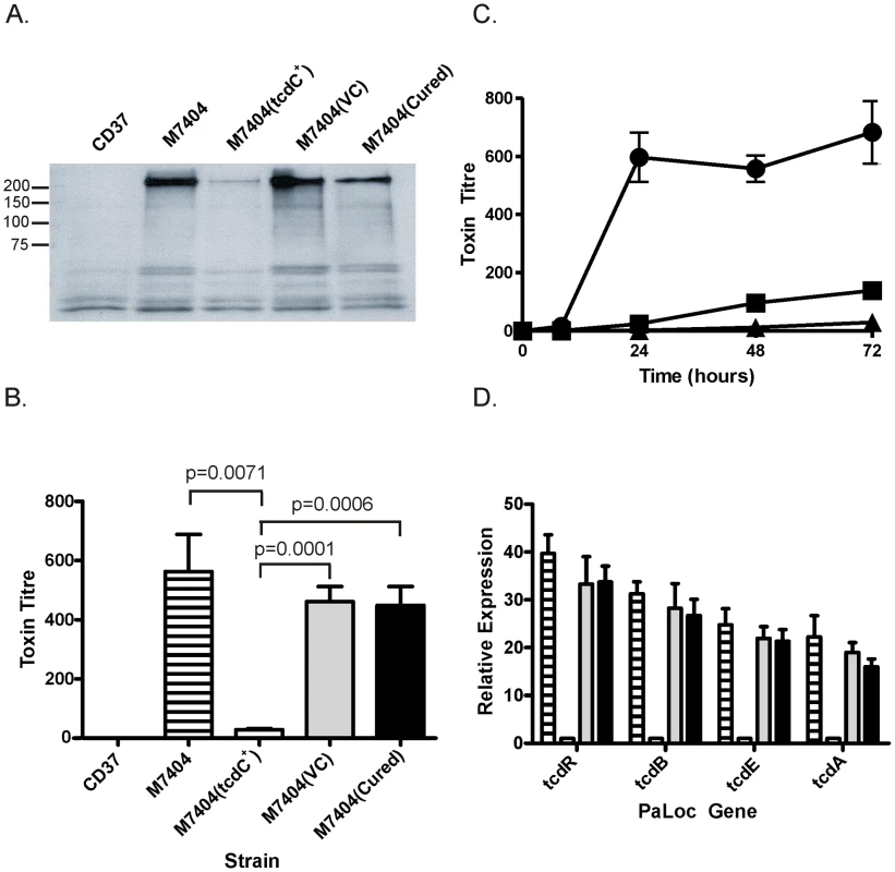
CD37 is the negative control strain; M7404 is the wild-type BI/NAPI/027 strain; M7404(tcdC+) is the wild-type strain carrying the tcdC expression vector pDLL17; M7404(VC) is the wild-type strain carrying shuttle plasmid pDLL4 and M7404(cured) is the M7404(tcdC+) strain cured of pDLL17. JIR8094 is a derivative of strain 630. (A) Western blot using toxin-A-specific antibodies. Size standards are shown (kDa) (B) Toxin cytotoxicity assays using Vero cells. Strains are as described above. Lined bars represent the wild-type strain M7404; white bars represent M7404(tcdC+); grey bars represent M7404(VC) and black bars represent M7404(cured). Data represent the mean ± s.e.m. (n = 3). (C) Time course of toxin production measured using Vero cell cytotoxicity assays. Strains are as described above and are represented as follows: M7404(tcdC+) (▪), M7404(VC) (•) and JIR8094 (▴). Note that CD37 was included in this analysis but displayed no toxin production; the line representing this strain is therefore not visible. Data represent the mean ± s.e.m. (n = 3). (D) PaLoc gene specific qRT-PCR. Bars correspond to strains as before and PaLoc genes are indicated. Data represent the mean fold-expression ± s.e.m. (n = 3), compared to the M7404(tcdC+) strain. Vero cell cytotoxicity assays, which predominantly measure toxin B activity [15], were then performed to quantitatively determine the effect of TcdC on toxin production from these strains. As previously observed with toxin A, the amount of toxin produced from strain M7404 was significantly reduced when functional TcdC was restored (Figure 2B). The amount of toxin produced in the TcdC-complemented strain was approximately 16–32-fold less, and therefore significantly lower (p = 0.0001; unpaired t-test, 95% confidence interval), than in the vector control-carrying M7404 derivative. There was, however, no significant difference in toxin activity levels between strains M7404, the vector control strain M7404(VC) or M7404(cured) (Figure 2B). A kinetic analysis of toxin production also clearly showed that the presence of TcdC delayed the onset of toxin production in M7404(tcdC+) in comparison to M7404 carrying the vector plasmid, mirroring the delayed toxin production observed from the tcdC+ 630-strain derivative JIR8094 (Figure 2C).
We also determined if TcdC-mediated repression of toxin production was at the transcriptional level and evaluated the effect of tcdC complementation on the expression of the other PaLoc-encoded genes, tcdR and tcdE. Quantitative real-time PCR (qRT-PCR) analysis using RNA extracted from the wild-type strain and its isogenic derivatives was performed to ascertain the relative transcription levels of the tcdA, tcdB, tcdR and tcdE genes. As shown in Figure 2D, an approximate 13 - and 23-fold reduction in tcdA- and tcdB-specific mRNA levels, respectively, in strain M7404(tcdC+) was observed compared to M7404. Similar observations were made for tcdR and tcdE expression levels, with 33-fold and 21-fold less tcdR and tcdE mRNA, respectively, in the tcdC-complemented strain compared to the wild-type. No significant differences in the expression levels of these four genes were detected when M7404, the vector-carrying derivative or the cured strain were compared. These data conclusively demonstrate that TcdC negatively regulates toxin production in C. difficile and show that repression occurs at the transcriptional level.
Reduction of the virulence of a BI/NAP1/027 isolate via complementation with TcdC
To define the role of TcdC in the virulence of a BI/NAP1/027 C. difficile isolate, female Golden Syrian hamsters were infected with spores of strain M7404 carrying either the vector control or the tcdC+ plasmid (n = 10 and n = 12, respectively). For comparative purposes, a group of hamsters (n = 14) was also infected with strain 630, a strain previously characterised as being less virulent than other clinical isolates [32]. Following infection, all C. difficile strains were found to be equally efficient at colonising the hamsters (data not shown). Infection of colonised hamsters was allowed to proceed and animals were monitored by telemetry. The end point of infection was achieved when the core body temperature of the hamsters dropped to 35°C. This parameter has previously been shown to be a reliable indicator of non-recoverable disease [33]. At this point, the animals were immediately culled for animal ethics reasons. Bacteria were then isolated from the culled hamsters, the bacterial load quantified and isolates subjected to MVLA analysis [33] to confirm that these isolates were the same strain as originally used for infection.
Hamsters infected with the M7404(tcdC+) derivative showed a significant delay (p = 0.0003; Logrank (Mantel-Cox) test; 95% confidence interval) in the mean time taken to reach non-recoverable disease (2370 minutes or 39.5 hours) in comparison to the vector-carrying M7404 group (M7404(VC)), with a mean time of 1869 minutes or 31.15 hours (Figure 3). In one of the hamsters colonised with the M7404(VC) strain, the time taken to reach the end point of infection was substantially longer than the other hamsters in this group (2814 minutes or 46.9 hours). This hamster was shown by statistical analysis (p = 0.0405; Grubbs test; 95% confidence interval) to be an outlier and was therefore excluded from the experimental analysis. Note that statistical significance would be retained upon inclusion of this outlier. Interestingly, whilst the mean time to the end point of infection in the strain 630 group of hamsters (2701 minutes or 45.02 hours) was significantly longer than that of hamsters infected with M7404(VC) (p = 0.0001; Logrank (Mantel-Cox) test; 95% confidence interval), there was no significant difference in the mean time taken to achieve non-recoverable disease in the 630 group compared to the M7404(tcdC+) derivative, indicating that the virulence of the TcdC-complemented strain was equivalent to that of strain 630.
Fig. 3. Virulence of C. difficile wild-type and tcdC-complemented strains in hamsters. 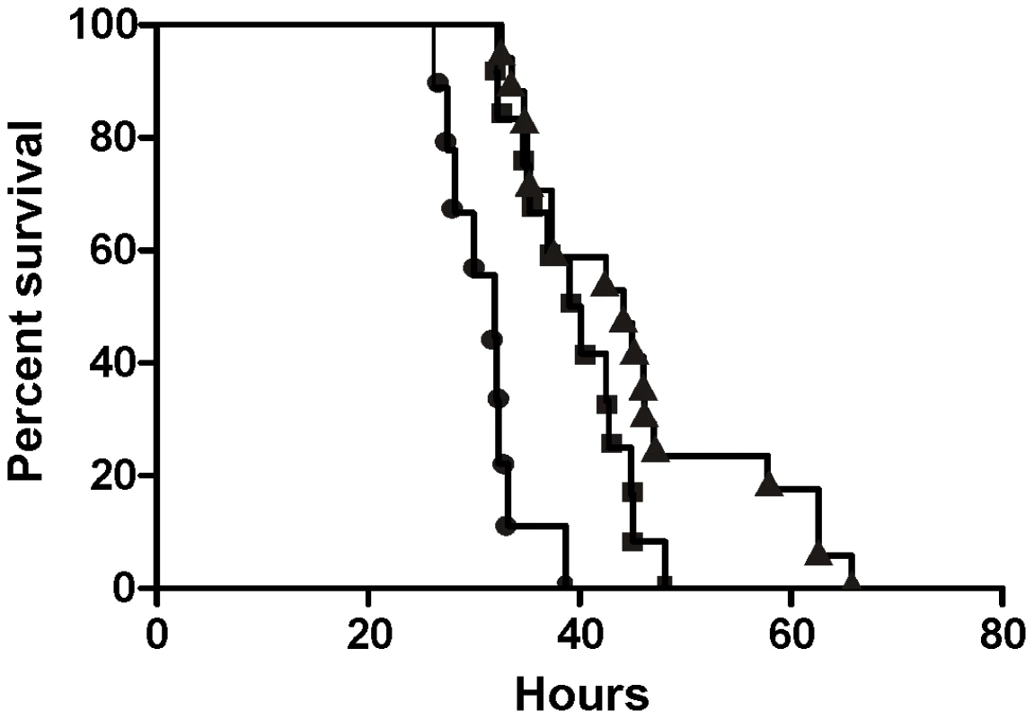
Kaplan-Meier survival curve demonstrating time from infection with C. difficile to death. M7404(VC), wild-type M7404 carrying shuttle plasmid pDLL4 (•); M7404(tcdC+), wild-type M7404 carrying tcdC expression plasmid pDLL17 (▪); and strain 630, a C. difficile isolate with known low virulence (▴). Hamsters were infected intragastrically with 10,000 spores from each strain; M7404(VC) (n = 9), M7404(tcdC+) (n = 12) and strain 630 (n = 14). It is apparent from these virulence experiments that the expression of TcdC in a BI/NAP1/027 isolate has an important effect on virulence, resulting in a significant delay in the time needed to reach non-recoverable disease. These data therefore provide compelling evidence that the naturally occurring mutation of tcdC in BI/NAP1/027 isolates is an important factor in the development of a hypervirulent phenotype by these strains.
TcdC status of clinical isolates does not predict toxin production level
The results presented here show that a BI/NAP1/027 strain complemented with tcdC is not as virulent as its isogenic vector-carrying control, suggesting that any C. difficile strain that acquires a null TcdC phenotype has the potential to develop a hypervirulent phenotype. Furthermore, phylogenetic studies have shown C. difficile to be a genetically diverse species, with disease-causing isolates seemingly arising from multiple lineages, suggesting that virulence in these strains may have evolved independently [4], [34]. The tcdC status of a diverse group of clinical isolates was therefore determined in parallel to the genetic studies described above. One hundred Australian clinical isolates were initially analysed by toxinotyping, a typing method which categorises strains according to variation in the PaLoc region; BI/NAP1/027 strains belong to toxinotype III [35]. Approximately 5% of these strains were found to belong to a toxinotype associated with a tcdC mutation. PCR and sequence analysis was then used to confirm the presence of tcdC mutations in each of these isolates. A single BI/NAP1/027 was identified in this survey, designated strain KI [36]. One other strain, DLL3053, was particularly interesting since it belonged to toxinotype group III but was not a BI/NAP1/027 isolate. This strain harboured the well documented single base pair deletion at nucleotide position 117 of tcdC, and the 18 base pair in-frame deletion from nucleotide 330 to 347 [37]. Two strains, DLL3054 and DLL3055, were toxinotype V strains with a nonsense mutation at nucleotide position 184 (C184T) and a 39 base pair deletion from nucleotides 341 to 379 [37] whereas strain DLL3056 was toxinotype XIV and possessed a nonsense mutation at nucleotide position 191 (C191A) and an in-frame 36 base pair deletion from nucleotide 300 to 336.
To examine the impact of tcdC mutations on toxin production in these clinical isolates, Vero cell cytotoxicity assays were performed. These assays, which predominantly detect toxin B, determine the relative amounts of toxin produced by each strain since the expression of toxins A and B is coordinately regulated [23], [31]. Strains JIR8094, a derivative of strain 630 [24], and VPI10463 [38], which both possess intact tcdC genes, were used as positive reference controls, and the PaLoc-negative strain CD37 was used as a negative control. Strains were grown in glucose-free medium since glucose has previously been shown to repress toxin production [23]. As shown in Figure 4A, the relative amount of toxin produced by the tcdC clinical isolates varied over approximately a 10-fold range. In agreement with the work of others [12], [22], these results show that the presence of mutations within tcdC was not directly correlated with high level toxin production. Strain DLL3053 for example, did not produce levels of toxin significantly different from that of strain JIR8094, which is considered to be a low toxin producer [39]. Of all the isolates with tcdC mutations, strain KI [36] produced the most toxin, and at significantly (p = 0.0406; unpaired t-test, 95% confidence interval) higher levels that were approximately 10-fold more than strain DLL3053 even though both strains have identical tcdC alleles.
Fig. 4. Comparative analysis of toxin production by naturally occurring tcdC clinical isolates. 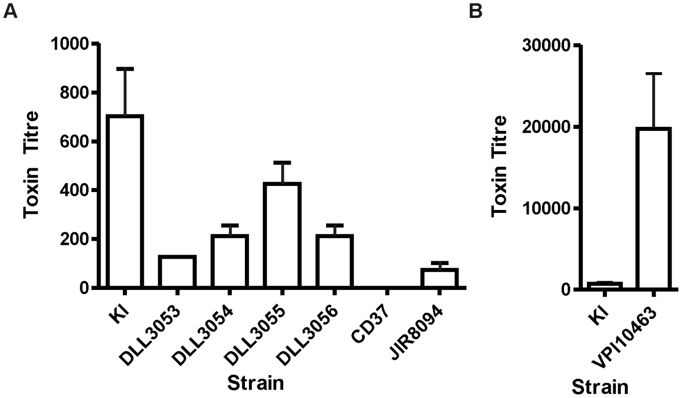
Vero cell cytotoxicity assays were used to determine toxin production levels. (A) Strain JIR8094 is a tcdC+ control strain, CD37 is a PaLoc-negative control strain and KI is an Australian BI/NAPI/027 isolate [36]. All other strains (DLL3053-DLL3056) are clinical isolates carrying naturally occurring tcdC mutations, collected from Australian hospitals. (B) Toxin production by the tcdC+ control strain VPI10463 is shown on a separate bar chart due to the much higher levels of toxin produced. For comparative purposes strain KI is represented on both bar charts. Data represent the mean±s.e.m. (n = 3). In comparison to the TcdC-positive reference strains, all of the tcdC-deficient clinical isolates produced more toxin than strain JIR8094 apart from strain DLL3053 (Figure 4A). Conversely, however, all strains produced significantly less toxin than VPI10463 (p = 0.0099–0.0202; unpaired t-test, 95% confidence interval), including the BI/NAP1/027 strain KI. VPI10463 produced over 100-fold more toxin than strains JIR8094 and DLL3053 and over 30-fold more than KI (Figure 4B). This observation is in agreement with recently published findings, which showed that VPI10463 produced significantly more toxin than other strains [12]; however, in that study, strains were grown using glucose-rich BHI medium so differential effects of glucose on toxin production by each strain could not be ruled out. In agreement with other studies [12], [22], [37], our data therefore suggest that the tcdC-status alone of C. difficile isolates is not an accurate predictor of high-level toxin production.
Discussion
The hypothesis that the naturally-occurring tcdC mutation in epidemic BI/NAP1/027 isolates contributes to hypervirulence is widely accepted, despite a lack of supportive experimental evidence. Indeed, the exact role of TcdC in the pathogenesis of C. difficile disease has remained controversial with conflicting findings reported in the literature [20]–[22]. As a result, several published studies have suggested that there is a need to assess isogenic tcdC strains in order to conclusively determine the role of this gene in the virulence of C. difficile [12], [22], [40]. We have now constructed such isogenic strains and compared them in an animal model. The results conclusively show that TcdC negatively regulates toxin production in C. difficile. Most importantly, complementation of the tcdC mutation in the BI/NAP1/027 epidemic isolate M7404 clearly showed that this mutation is an important factor in the development of hypervirulence by this strain since the genetic complementation of tcdC reduced virulence in comparison to the wild-type strain.
To elucidate the role of TcdC in hypervirulence, it was necessary to construct an isogenic panel of BI/NAP1/027 strains that were identical except for the presence or absence of the wild-type tcdC gene. Despite the publication of studies describing the successful transfer of plasmids into the BI/NAP1/027 isolate R20291 [25], [26], [41], this group of strains has remained difficult to work with at the molecular genetic level. As such, a new system that utilised the conjugation apparatus of Tn916 was developed in this study and used successfully to genetically manipulate a number of clinically relevant isolates, including a BI/NAP1/027 strain of C. difficile. Tn916 is a broad host-range conjugative transposon that was recently used to transfer plasmids into genetically intractable strains of Enterococcus faecium [28] and has been shown to transfer into C. difficile [42], [43]. C. perfringens was chosen for use as a donor strain in anticipation that it may be more proficient for the transfer of plasmids into C. difficile in comparison to the more distantly related E. coli. The addition of oriTTn916 onto the shuttle vector pMTL9361Cm facilitated the efficient transfer of this plasmid into strain M7404 from a Tn916-carrying C. perfringens strain. Furthermore, this system has been successfully used to transfer shuttle plasmids into every C. difficile isolate tested so far (Table S1). Most importantly, this new technology facilitated the complementation of the tcdC mutation in strain M7404 enabling the role of TcdC in the virulence of BI/NAP1/027 strains of C. difficile to be investigated.
Previous in vitro studies have shown that TcdC is able to sequester the TcdR sigma factor, preventing its association with core RNA polymerase and blocking toxin gene expression [20]. These experiments suggested that TcdC was important in the regulation of toxin production by C. difficile, but the in vivo role of this protein was not determined. Conversely, several studies on C. difficile clinical isolates [22], [37] showed that the absence of a functional tcdC gene was not an accurate predictor of high level toxin production or increased disease severity, indicating that TcdC may not play an important role in virulence in these strains [22], [37]. The analysis of Australian clinical isolates in the present study is in accordance with these latter studies in that isolates with naturally occurring tcdC mutations were found to produce toxin at a range of different levels that were not necessarily high. However, since these strains, and those in the other studies [22], [37], are not isogenic it is not possible to draw conclusions about the importance of tcdC in the context of toxin yield or virulence. By contrast, the isogenic tcdC strains studied here clearly show that TcdC is a negative regulator of toxin production since the tcdC complemented BI/NAP1/027 C. difficile strain produced significantly less toxin A and B than the non-complemented control strains. The finding that TcdC-status is not correlated with toxin production in clinical isolates highlights the limitation of accurately assigning gene function by studying non-isogenic strains, particularly in a highly heterogeneous species such as C. difficile. In this context, it might be of interest to study the function of tcdC in isogenic strains generated in a different genetic background such as a ribotype 078 isolate.
Analysis of PaLoc gene expression by qRT-PCR demonstrated that TcdC exerts regulatory control of toxin production at the transcriptional level, and this is in keeping with its proposed role as an anti-sigma factor [20]. The observation that the expression of tcdR and tcdE is reduced in the tcdC-complemented strain, together with tcdA and tcdB, is probably because of autoregulation of tcdR since TcdR upregulates its own expression and that of the other PaLoc genes [23].
The virulence of strain M7404 was reduced upon complementation of tcdC, clearly demonstrating that the tcdC mutation in BI/NAP1/027 strains has a significant impact on virulence and is likely to be an important factor in the development of hypervirulence by these strains. Surprisingly, the virulence of M7404(tcdC+) was found to be equivalent to that of strain 630, which has been shown in other studies to be reduced in virulence in comparison to other isolates, including three other BI-type strains [32]. These findings have important implications for C. difficile virulence since they suggest that strains carrying tcdC mutations have the inherent potential to develop hypervirulence. The recent emergence of a new class of hypervirulent strains, ribotype 078 [44], may be one such example. These isolates encode a non-functional TcdC protein [37], produce significantly more toxin than non-epidemic strains, are associated with more severe disease as well as higher rates of mortality and are increasingly being identified as the causative agent of CDI [44], [45].
Although these experiments show that TcdC-status alone can modulate virulence it is probable that multiple factors working synergistically are necessary for the development of hypervirulence in the BI/NAP1/027 strains. It is likely that the accumulation of multiple genetic changes in addition to the tcdC mutation has enabled BI/NAP1/027 strains to become the predominant disease-causing isolates in numerous countries. Of particular importance might be variations in the functional activity of the encoded toxins since these isolates were recently shown to produce a toxin B that shows variation across the C-terminal receptor binding domain of the protein [46], resulting in more potent activity across a wider range of cell lines in comparison to toxin B from the historical, non-epidemic strain 630 [4]. Furthermore, using the zebrafish embryo model of intoxication, the BI/NAP1/027 toxin B was recently shown to have pronounced in vivo cytotoxic activity in comparison to toxin B from VPI10463, another historical non-epidemic isolate, with greater tissue tropism and more extensive tissue destruction observed [47]. Since toxin B is thought to be one of the major virulence factors of C. difficile [15], [16] these observations suggest that TcdB variations might play an important role in the hypervirulent phenotype.
There are other factors that may influence C. difficile hypervirulence. The BI/NAP1/027 strains encode an additional toxin known as binary toxin or CDT [2]. The role of this toxin in CDI remains to be elucidated but a recent study showed that CDT induces the formation of microtubule-based protrusions on the host cell surface thereby increasing C. difficile adherence to epithelial cells. Moreover, intestinal colonisation of gnotobiotic mice with a BI/NAP1/027 C. difficile strain was significantly reduced in mice treated with CDT-neutralising antibodies in comparison to control mice [48]. These findings suggest that CDT may be an important colonisation factor, enhancing the ability of BI/NAP1/027 strains to initiate infection as well as causing adjunctive tissue damage during later stages of infection, potentially leading to more severe disease. Many BI/NAP1/027 strains are also more proficient at sporulation than non-epidemic C. difficile strains [12], [49]. C. difficile spores are highly infectious [50] and play a critical role in the transmission of CDI and perhaps in disease relapse, which is a serious problem in patients with CDI [51]. In this context, enhanced sporulation is ostensibly an important adaptation by BI/NAP1/027 isolates, which would result in larger numbers of spores being shed from infected patients and an increased environmental spore load, ultimately leading to higher transmission rates. Finally, the development of fluoroquinolone resistance, in particular to moxifloxacin and gatifloxacin, is unquestionably a major factor in epidemics caused by BI/NAP1/027 strains [3], [11]. In this regard, the hypothetical co-evolution of enhanced virulence traits and antibiotic resistance in C. difficile mirrors trends seen with other significant nosocomial pathogens such as Methicillin Resistant Staphylococcus aureus (MRSA) [52] and Vancomycin Resistant Enterococcus (VRE) species [53]. In summary, it is clear that our findings represent an important breakthrough in our understanding of the development of hypervirulence in prevailing C. difficile isolates and will provide a significant reference point for future studies on epidemic strains and their control.
Materials and Methods
Ethics statement
This study was carried out in strict accordance with the recommendations in the United Kingdoms Home Office Animals (Scientific Procedures) Act of 1986 which outlines the regulation of the use of laboratory animals for the use of animals in scientific procedures. The experiments described were subject to approval by the University of Glasgow Ethics Committee and by a designated Home Office Inspector (Project Number 60/4218). All experiments were subject to the 3 R consideration (refine, reduce and replace) and all efforts were made to minimize suffering.
Bacterial strains and growth conditions
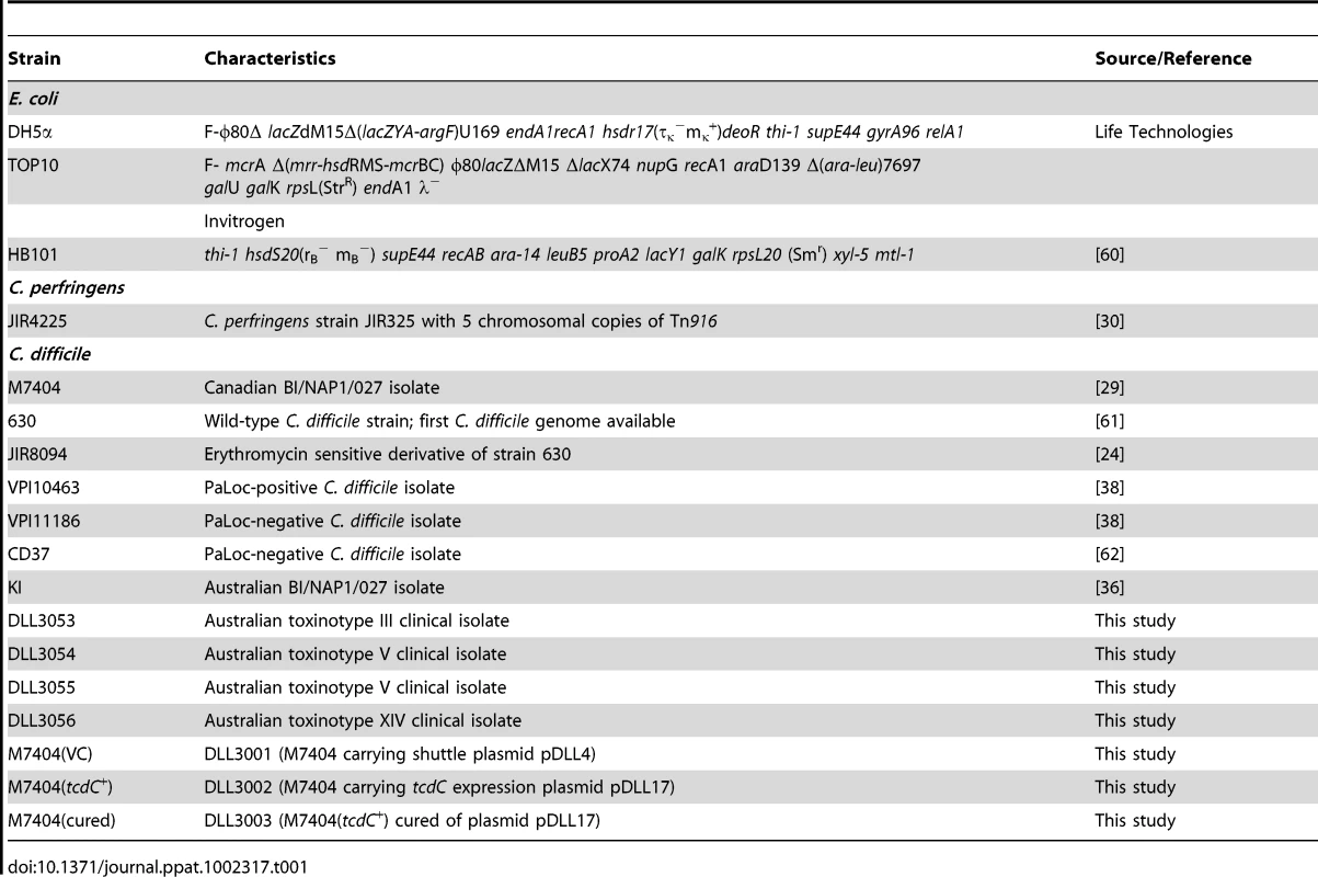
The characteristics and origins of all recombinant strains and plasmids are shown in Table 1 and Table S2, respectively. All bacteriological culture media were obtained from Oxoid. C. difficile strains were cultured in BHIS [54] or TY medium [15], unless otherwise stated, in an atmosphere of 10% H2, 10% CO2, and 80% N2 at 37°C in a Coy anaerobic chamber. Escherichia coli was cultured in 2×YT medium aerobically at 37°C, with shaking for broth cultures. All antibiotics were purchased from Sigma-Aldrich and were used at the following concentrations: cycloserine (Cs, 250 µg/ml), cefoxitin (Cf, 8 µg/ml), thiamphenicol (Tm, 10 µg/ml) or tetracycline (Tc, 10 µg/ml), chloramphenicol (Cm, 25 µg/ml).
Molecular biology and PCR techniques
Plasmid DNA was isolated using a QIAprep spin miniprep kit (Qiagen). Genomic DNA was prepared using a DNeasy tissue kit (Qiagen). Standard methods for the digestion, modification, ligation, and analysis of plasmid and genomic DNA were used [55]. Nucleotide sequence analysis was carried out using a PRISM BigDye Terminator cycle sequencing kit (Applied Biosystems) and detection was performed by Micromon at Monash University. Oligonucleotide primer sequences are listed below. Unless otherwise stated, all PCR experiments were carried out with Phusion DNA polymerase (New England Biolabs) and the 2× Failsafe PCR buffer E (Epicentre) according to the manufacturer's instructions.
Construction of recombinant plasmids
For construction of the Tn916 transferrable clostridial shuttle vector, PCR was performed using primers DLP33 (5′-GAATTCGCCCTTTTTTATACTCCCCTTG-3′) and DLP34 (5′-GAATTCGCCCTCAAAGGACGAATATGTCGC-3′) and chromosomal DNA extracted from Clostridium perfringens strain JIR4225 [30]. The resulting 700 bp DNA fragment, which contained the oriT region of Tn916, was TOPO-cloned into pCR-Blunt II-TOPO according to the manufacturer's instructions (Invitrogen). The fragment was then excised from pCR-Blunt II-TOPO using EcoRI and cloned into the equivalent sites of plasmid pMTL9361Cm [29], resulting in plasmid pDLL4.
For construction of the tcdC-carrying plasmid, PCR was performed using primers DLP35 (5′-CTGCAGCCACCTCTAAATCACTGAGTCACTTAATTAC-3′) and DLP36 (5′-CTGCAGAGCCTTGTAACTGTTTATTTGC-3′) and C. difficile strain VPI10463 genomic DNA in order to amplify a 1085 bp fragment encompassing the tcdC gene and upstream region. This fragment was then TOPO-cloned into pCR-Blunt II-TOPO, before being excised with PstI and subcloned into the equivalent site of plasmid pDLL4, resulting in the final construct pDLL17.
Transfer of plasmid DNA into C. difficile by conjugation
The conjugation procedure utilising E. coli HB101(pVS520) as the conjugative donor was carried out as previously described [29]. Recombinant plasmids were introduced into C. perfringens strain JIR4225 as before [56]. Conjugations utilising C. perfringens JIR4225 were then performed as follows: separate 90 ml BHIS broth cultures were inoculated with 1 ml aliquots from an overnight C. difficile recipient strain or C. perfringens donor strain starter culture and grown to mid-exponential phase. Approximately 1 ml was removed from each culture, mixed and centrifuged. The cell pellet was then resuspended in phosphate-buffered saline (PBS), spread onto a BHIS agar plate and incubated overnight at 37°C. Bacterial growth was harvested in sterile PBS before being spread onto BHIS agar supplemented with thiamphenicol and incubated overnight as before. Bacterial growth was again harvested with PBS and dilutions spread onto BHIS agar supplemented with cefoxitin and thiamphenicol or tetracycline, and the plates incubated under anaerobic conditions for 24 to 72 h.
Toxin A-specific Western blots
The toxins were partially purified by ammonium sulphate precipitation from culture supernatants harvested after growth for 72 hours and toxin A was then detected by Western blotting as described previously [15].
TcdC-specific Western blots
For non-quantitative TcdC-specific Western Blots, crude extracts of C. difficile were prepared by sonication of samples taken from cultures that had been grown for 12 hours under anaerobic conditions. The crude extracts from each strain were then subjected to electrophoresis in a 15% SDS-PAGE gel and transferred to a nitrocellulose membrane using standard methods [55]. Membranes were treated with anti-TcdC antibody [57] and detected following treatment with goat anti-mouse IgG-alkaline phosphatase conjugated secondary antibody using standard procedures. For quantitative TcdC-specific Western blots, cultures of C. difficile VPI10463 and C. difficile M7404(tcdC+) were grown under anaerobic conditions and samples were removed every 4 hours for 24 or 28 hours respectively. Each sample was normalized to an optical density (600 nm) of 0.9 prior to lysis, to ensure that the same number of cells was present. Lysates were then prepared by sonication prior to SDS-PAGE gel electrophoresis, transfer and detection, as described above. Following detection, the amount of TcdC in each lysate was quantified by densitometric analysis, using purified recombinant TcdC protein (rTcdC) as the standard and the ImageJ software package, according to published methods [58].
Vero cell cytotoxicity assays
Toxin B was detected in C. difficile culture supernatants harvested after growth for 72 hours by Vero cell cytotoxicity assays as described previously [15], except that each well was seeded with 1×10⁵ cells.
RNA extraction and reverse transcription
Total RNA was extracted from C. difficile cultures grown for 12 h in TY media. Reverse transcription was performed using AMV Reverse Transcriptase (Promega) using random hexamer oligonucleotides primers and 2 µg template RNA. The cDNA samples were then purified using Qiaquick Columns (Qiagen).
qRT-PCR assay design and real time PCR
PaLoc gene specific primers were designed using Primer 3 software (Geneious Software). qRT-PCR was performed using an AB7300 real-time PCR instrument (Applied Biosystems). Reactions were carried out using the FastStart Universal SYBR Green Master Mix (Roche) with 40 ng of cDNA as template. Standard curves were generated for each primer pair using C. difficile genomic DNA, and melt curve analysis was performed following each qRT-PCR reaction to verify amplification specificity. Samples were normalised using the C. difficile rrnA gene.
Preparation of spores for animal infection
Spores were prepared from C. difficile cultures grown in 500 ml of BHI broth. Cultures were pelleted by centrifugation for 10 mins and re-suspended in 50% ethanol. The material was then vortexed every 10 min for 1 h before centrifugation for 10 mins. The pellet was then treated with 1% Sarkosyl in PBS for 1 h at room temperature and again pelleted by centrifugation, followed by incubation overnight at 37°C with lysozyme (10 mg/ml) in 125 mM Tris-HCl buffer (pH 8.0). The sample was treated in a sonicating water bath (3 pulses of 3 min each; 1510 Branson) before centrifugation through a 50% sucrose gradient for 20 mins. The pellet was incubated in 2 ml of PBS containing 200 mM EDTA, 300 ng/ml proteinase K and 1% Sarkosyl for 30 mins at 37°C before centrifugation through a 50% sucrose gradient for 20 mins. The final pellet was then washed twice in sterile distilled water before finally being resuspended in 1 ml of sterile water. Spore preparations were stored at −80°C prior to use.
Hamster experiments
Female Golden Syrian hamsters purchased from Harlan Olac UK were used for all animal experiments. Telemetry chips (Vitalview Emitter) were inserted by laparotomy into the body cavity of the animals at least 3 weeks before infection with C. difficile. Animal experiments were then carried out as described previously [33], except that animals received 1×10⁴ spores of C. difficile. Animals were culled when core body temperature dropped below 35°C. This study was carried out in strict accordance with the recommendations in the United Kingdoms Home Office Animals (Scientific Procedures) Act of 1986 which outlines the regulation of the use of laboratory animals for the use of animals in scientific procedures. The experiments described were subject to approval by the University of Glasgow Ethics Committee and by a designated Home Office Inspector (Project Number 60/4218). All experiments were subject to the 3 R consideration (refine, reduce and replace) and all efforts were made to minimize suffering.
Quantification of bacterial load
To estimate colonisation, hamsters were sacrificed and the gut region from the caecum to the anus removed. The tissues were homogenised in PBS using a Stomacher and viable counts were performed on the homogenate as described previously [33].
Confirmation of infecting strains
To confirm that the bacteria isolated from the hamster were the same strain as originally used for infection, genomic DNA was isolated and subjected to MVLA as described previously [59]. Plasmid rescue was performed as previously described [29] followed by restriction digest analysis to confirm plasmid integrity.
Supporting Information
Zdroje
1. BorrielloSP 1998 Pathogenesis of Clostridium difficile infection. J Antimicrob Chemother 41 13 19
2. WarnyMPepinJFangAKillgoreGThompsonA 2005 Toxin production by an emerging strain of Clostridium difficile associated with outbreaks of severe disease in North America and Europe. Lancet 366 1079 1084
3. McDonaldLCKillgoreGEThompsonAOwensRCKazakovaSV 2005 An epidemic, toxin gene–variant strain of Clostridium difficile. N Engl J Med 353 2433 2441
4. StablerRAHeMDawsonLMartinMValienteE 2009 Comparative genome and phenotypic analysis of Clostridium difficile 027 strains provides insight into the evolution of a hypervirulent bacterium. Genome Biol 10 R102
5. LooVGPoirierLMillerMAOughtonMLibmanMD 2005 A predominantly clonal multi-institutional outbreak of Clostridium difficile-associated diarrhea with high morbidity and mortality. N Engl J Med 353 2442 2449
6. MutoCAPokrywkaMShuttKMendelsohnABNouriK 2005 A large outbreak of Clostridium difficile-associated disease with an unexpected proportion of deaths and colectomies at a teaching hospital following increased fluoroquinolone use. Infect Control Hosp Epidemiol 26 273 280
7. KuijperEJCoignardBTullP 2006 Emergence of Clostridium difficile-associated disease in North America and Europe. Clin Microbiol Infect 12 Suppl 6 2 18
8. RouphaelNGO'DonnellJABhatnagarJLewisFPolgreenPM 2008 Clostridium difficile-associated diarrhea: an emerging threat to pregnant women. Am J Obstet Gynecol 198 635 e631 636
9. ZilberbergMDTillotsonGSMcDonaldC 2010 Clostridium difficile infections among hospitalized children, United States, 1997–2006. Emerg Infect Dis 16 604 609
10. BakerSSFadenHSayejWPatelRBakerRD 2010 Increasing Incidence of Community-Associated Atypical Clostridium difficile Disease in Children. Clin Pediatr (Phila) 49 644 647
11. RileyTV 2009 Is Clostridium difficile a threat to Australia's biosecurity? Med J Aust 190 661 662
12. MerriganMVenugopalAMallozziMRoxasBViswanathanVK 2010 Human Hypervirulent Clostridium difficile Strains Exhibit Increased Sporulation as Well as Robust Toxin Production. J Bacteriol 192 4904 4911
13. SebaihiaMWrenBWMullanyPFairweatherNFMintonN 2006 The multidrug-resistant human pathogen Clostridium difficile has a highly mobile, mosaic genome. Nat Genet 38 779 786
14. JustISelzerJWilmMvon Eichel-StreiberCMannM 1995 Glucosylation of Rho proteins by Clostridium difficile toxin B. Nature 375 500 503
15. LyrasDO'ConnorJRHowarthPKSambolSPCarterGP 2009 Toxin B is essential for virulence of Clostridium difficile. Nature 458 1176 1179
16. KuehneSACartmanSTHeapJTKellyMLCockayneA 2010 The role of toxin A and toxin B in Clostridium difficile infection. Nature 467 711 713
17. BraunVHundsbergerTLeukelPSauerbornMEichel-StreiberCv 1996 Definition of the single integration site of the pathogenicity locus in Clostridium difficile. Gene 181 29 38
18. ManiNDupuyB 2001 Regulation of toxin synthesis in Clostridium difficile by an alternative RNA polymerase sigma factor. Proc Natl Acad Sci U S A 98 5844 5849
19. TanKSWeeBYSongKP 2001 Evidence for holin function of tcdE gene in the pathogenicity of Clostridium difficile. J Med Microbiol 50 613 619
20. MatamourosSEnglandPDupuyB 2007 Clostridium difficile toxin expression is inhibited by the novel regulator TcdC. Mol Microbiol 64 1274 1288
21. DupuyBGovindRAntunesAMatamourosS 2008 Clostridium difficile toxin synthesis is negatively regulated by TcdC. J Med Microbiol 57 685 689
22. MurrayRBoydDLevettPNMulveyMRAlfaMJ 2009 Truncation in the tcdC region of the Clostridium difficile PathLoc of clinical isolates does not predict increased biological activity of Toxin B or Toxin A. BMC Infect Dis 9 103
23. ManiNLyrasDBarrosoLHowarthPWilkinsT 2002 Environmental response and autoregulation of Clostridium difficile TxeR, a sigma factor for toxin gene expression. J Bacteriol 184 5971 5978
24. O'ConnorJRLyrasDFarrowKAAdamsVPowellDR 2006 Construction and analysis of chromosomal Clostridium difficile mutants. Mol Microbiol 61 1335 1351
25. BurnsDAHeapJTMintonNP 2010 SleC is essential for germination of Clostridium difficile spores in nutrient-rich medium supplemented with the bile salt taurocholate. J Bacteriol 192 657 664
26. CartmanSTMintonNP 2010 A mariner-based transposon system for in vivo random mutagenesis of Clostridium difficile. Appl Environ Microbiol 76 1103 1109
27. HeapJTKuehneSAEhsaanMCartmanSTCooksleyCM 2010 The ClosTron: Mutagenesis in Clostridium refined and streamlined. J Microbiol Methods 80 49 55
28. NallapareddySRSinghKVMurrayBE 2006 Construction of improved temperature-sensitive and mobilizable vectors and their use for constructing mutations in the adhesin-encoding acm gene of poorly transformable clinical Enterococcus faecium strains. Appl Environ Microbiol 72 334 345
29. CarterGPLyrasDAllenDLMackinKEHowarthPM 2007 Binary toxin production in Clostridium difficile is regulated by CdtR, a LytTR family response regulator. J Bacteriol 189 7290 7301
30. AwadMMRoodJI 1997 Isolation of a-toxin, q-toxin and k-toxin mutants of Clostridium perfringens by Tn916 mutagenesis. Microb Pathogen 22 275 284
31. HundsbergerTBraunVWeidmannMLeukelPSauerbornM 1997 Transcription analysis of the genes tcdA-E of the pathogenicity locus of Clostridium difficile. European J Bioch 244 735 742
32. RazaqNSambolSNagaroKZukowskiWCheknisA 2007 Infection of hamsters with historical and epidemic BI types of Clostridium difficile. J Infect Dis 196 1813 1819
33. GouldingDThompsonHEmersonJFairweatherNFDouganG 2009 Distinctive profiles of infection and pathology in hamsters infected with Clostridium difficile strains 630 and B1. Infect Immun 77 5478 5485
34. HeMSebaihiaMLawleyTDStablerRADawsonLF 2010 Evolutionary dynamics of Clostridium difficile over short and long time scales. Proc Natl Acad Sci U S A 107 7527 7532
35. RupnikMAvesaniVJancMvon Eichel-StreiberCDelmeeM 1998 A novel toxinotyping scheme and correlation of toxinotypes with serogroups of Clostridium difficile isolates. J Clin Microbiol 36 2240 2247
36. RichardsMKnoxJElliottBMackinKLyrasD 2011 Severe infection with Clostridium difficile PCR ribotype 027 acquired in Melbourne, Australia. Med J Australia 194 369 371
37. CurrySRMarshJWMutoCAO'LearyMMPasculleAW 2007 tcdC genotypes associated with severe TcdC truncation in an epidemic clone and other strains of Clostridium difficile. J Clin Microbiol 45 215 221
38. LyerlyDMBarrosoLAWilkinsTDDepitreCCorthierG 1992 Characterization of a toxin A-negative, toxin B-positive strain of Clostridium difficile. Infect Immun 60 4633 4639
39. CarterGPRoodJILyrasD 2010 The role of toxin A and toxin B in Clostridium difficile-associated disease: Past and present perspectives. Gut Microbes 1 58 64
40. O'ConnorJRJohnsonSGerdingDN 2009 Clostridium difficile infection caused by the epidemic BI/NAP1/027 strain. Gastroenterology 136 1913 1924
41. HeapJTPenningtonOJCartmanSTCarterGPNP.M 2007 The ClosTron: a universal gene knock-out system for the genus Clostridium. J Microbiol Meth 70 452 464
42. MullanyPWilksMTabaqchaliS 1991 Transfer of Tn916 and Tn916DE into Clostridium difficile: demonstration of a hot-spot for these elemnets in the C. difficile genome. FEMS Microbiol Lett 79 191 194
43. MintonNCarterGHerbertMO'KeeffeTPurdyD 2004 The development of Clostridium difficile genetic systems. Anaerobe 10 75 84
44. GoorhuisADebastSBvan LeengoedLAHarmanusCNotermansDW 2008 Clostridium difficile PCR ribotype 078: an emerging strain in humans and in pigs? J Clin Microbiol 46 1157; author reply 1158
45. DawsonLFValienteEWrenBW 2009 Clostridium difficile–a continually evolving and problematic pathogen. Infect Genet Evol 9 1410 1417
46. StablerRADawsonLFPhuaLTWrenBW 2008 Comparative analysis of BI/NAP1/027 hypervirulent strains reveals novel toxin B-encoding gene (tcdB) sequences. J Med Microbiol 57 771 775
47. LanisJMBaruaSBallardJD 2010 Variations in TcdB activity and the hypervirulence of emerging strains of Clostridium difficile. PLoS Pathog 6 e1001061
48. SchwanCStecherBTzivelekidisTvan HamMRohdeM 2009 Clostridium difficile toxin CDT induces formation of microtubule-based protrusions and increases adherence of bacteria. PLoS Pathog 5 e1000626
49. AkerlundTPerssonIUnemoMNorenTSvenungssonB 2008 Increased sporulation rate of epidemic Clostridium difficile Type 027/NAP1. J Clin Microbiol 46 1530 1533
50. LawleyTDClareSWalkerAWGouldingDStablerRA 2009 Antibiotic treatment of Clostridium difficile carrier mice triggers a supershedder state, spore-mediated transmission, and severe disease in immunocompromised hosts. Infect Immun 77 3661 3669
51. JohnsonS 2009 Recurrent Clostridium difficile infection: a review of risk factors, treatments, and outcomes. J Infect 58 403 410
52. LindsayJA 2010 Genomic variation and evolution of Staphylococcus aureus. Int J Med Microbiol 300 98 103
53. BontenMJWillemsRWeinsteinRA 2001 Vancomycin-resistant enterococci: why are they here, and where do they come from? Lancet Infect Dis 1 314 325
54. SmithCJMarkowitzSMMacrinaFL 1981 Transferable tetracycline resistance in Clostridium difficile. Antimicrob Agents and Chemother 19 997 1003
55. SambrookJRussellDW 2001 Molecular Cloning: A Laboratory Manual Cold Spring Harbor, NY Cold Spring Harbor Laboratory Press
56. AwadMMBryantAEStevensDLRoodJI 1995 Virulence studies on chromosomal a-toxin and q-toxin mutants constructed by allelic exchange provide genetic evidence for the essential role of a-toxin in Clostridium perfringens-mediated gas gangrene. Mol Microbiol 15 191 202
57. GovindRVediyappanGRolfeRDFralickJA 2006 Evidence that Clostridium difficile TcdC is a membrane-associated protein. J Bacteriol 188 3716 3720
58. AbramoffMDMagelhaesPJRamSJ 2004 Image Processing with ImageJ. Biophotonics Internatl 11 36 42
59. MarshJWO'LearyMMShuttKAPasculleAWJohnsonS 2006 Multilocus variable-number tandem-repeat analysis for investigation of Clostridium difficile transmission in Hospitals. J Clin Microbiol 44 2558 2566
60. BoyerHWRoulland-DussoixD 1969 A complementation analysis of the restriction and modification of DNA in Escherichia coli. J Mol Biol 41 459 472
61. WustJSullivanNMHardeggerUWilkinsTD 1982 Investigation of an outbreak of antibiotic-associated colitis by various typing methods. J Clin Microbiol 16 1096 1101
62. MullanyPWilksMLambIClaytonCWrenB 1990 Genetic analysis of a tetracycline resistance element from Clostridium difficile and its conjugal transfer to and from Bacillus subtilis. J Gen Microbiol 136 1343 1349
Štítky
Hygiena a epidemiológia Infekčné lekárstvo Laboratórium
Článek Quorum Sensing in Fungi: Q&AČlánek Blood Feeding and Insulin-like Peptide 3 Stimulate Proliferation of Hemocytes in the MosquitoČlánek The DEAD-box RNA Helicase DDX6 is Required for Efficient Encapsidation of a Retroviral GenomeČlánek A Phenome-Based Functional Analysis of Transcription Factors in the Cereal Head Blight Fungus,Článek A Wide Extent of Inter-Strain Diversity in Virulent and Vaccine Strains of AlphaherpesvirusesČlánek Critical Roles for LIGHT and Its Receptors in Generating T Cell-Mediated Immunity during InfectionČlánek Frequent and Recent Human Acquisition of Simian Foamy Viruses Through Apes' Bites in Central Africa
Článok vyšiel v časopisePLOS Pathogens
Najčítanejšie tento týždeň
2011 Číslo 10- Parazitičtí červi v terapii Crohnovy choroby a dalších zánětlivých autoimunitních onemocnění
- Očkování proti virové hemoragické horečce Ebola experimentální vakcínou rVSVDG-ZEBOV-GP
- Koronavirus hýbe světem: Víte jak se chránit a jak postupovat v případě podezření?
-
Všetky články tohto čísla
- Quorum Sensing in Fungi: Q&A
- Discovery of an Ebolavirus-Like Filovirus in Europe
- Toll-like Receptor 7 Controls the Anti-Retroviral Germinal Center Response
- Tubule-Guided Cell-to-Cell Movement of a Plant Virus Requires Class XI Myosin Motors
- Herpesvirus Telomerase RNA (vTR) with a Mutated Template Sequence Abrogates Herpesvirus-Induced Lymphomagenesis
- Mitochondrial Peroxiredoxin Plays a Crucial Peroxidase-Unrelated Role during Infection: Insight into Its Novel Chaperone Activity
- Sustained CD8+ T Cell Memory Inflation after Infection with a Single-Cycle Cytomegalovirus
- Novel Mouse Xenograft Models Reveal a Critical Role of CD4 T Cells in the Proliferation of EBV-Infected T and NK Cells
- Toll-8/Tollo Negatively Regulates Antimicrobial Response in the Respiratory Epithelium
- Exhausted Cytotoxic Control of Epstein-Barr Virus in Human Lupus
- Structural and Functional Analysis of Laninamivir and its Octanoate Prodrug Reveals Group Specific Mechanisms for Influenza NA Inhibition
- Infection Drives IL-17-Mediated Neutrophilic Allergic Airways Disease
- Blood Feeding and Insulin-like Peptide 3 Stimulate Proliferation of Hemocytes in the Mosquito
- HIV-1 Replication in the Central Nervous System Occurs in Two Distinct Cell Types
- Deep Molecular Characterization of HIV-1 Dynamics under Suppressive HAART
- Fitness Landscape of Antibiotic Tolerance in Biofilms
- The DEAD-box RNA Helicase DDX6 is Required for Efficient Encapsidation of a Retroviral Genome
- Preventing Sepsis through the Inhibition of Its Agglutination in Blood
- A Phenome-Based Functional Analysis of Transcription Factors in the Cereal Head Blight Fungus,
- IFITM3 Inhibits Influenza A Virus Infection by Preventing Cytosolic Entry
- Targeting Cattle-Borne Zoonoses and Cattle Pathogens Using a Novel Trypanosomatid-Based Delivery System
- A Wide Extent of Inter-Strain Diversity in Virulent and Vaccine Strains of Alphaherpesviruses
- Coordinated Destruction of Cellular Messages in Translation Complexes by the Gammaherpesvirus Host Shutoff Factor and the Mammalian Exonuclease Xrn1
- Signal Transduction through CsrRS Confers an Invasive Phenotype in Group A
- Biochemical and Structural Insights into the Mechanisms of SARS Coronavirus RNA Ribose 2′-O-Methylation by nsp16/nsp10 Protein Complex
- Histone Deacetylase 8 Is Required for Centrosome Cohesion and Influenza A Virus Entry
- Severe Acute Respiratory Syndrome Coronavirus Envelope Protein Regulates Cell Stress Response and Apoptosis
- Co-opts the FGF2 Signaling Pathway to Enhance Infection
- IRAK-2 Regulates IL-1-Mediated Pathogenic Th17 Cell Development in Helminthic Infection
- Trafficking of Hepatitis C Virus Core Protein during Virus Particle Assembly
- The Anti-interferon Activity of Conserved Viral dUTPase ORF54 is Essential for an Effective MHV-68 Infection
- A Viral Nuclear Noncoding RNA Binds Re-localized Poly(A) Binding Protein and Is Required for Late KSHV Gene Expression
- Suppression of Methylation-Mediated Transcriptional Gene Silencing by βC1-SAHH Protein Interaction during Geminivirus-Betasatellite Infection
- ISG15 Is Critical in the Control of Chikungunya Virus Infection Independent of UbE1L Mediated Conjugation
- Non-Hematopoietic Cells in Lymph Nodes Drive Memory CD8 T Cell Inflation during Murine Cytomegalovirus Infection
- RNA Polymerase II Stalling Promotes Nucleosome Occlusion and pTEFb Recruitment to Drive Immortalization by Epstein-Barr Virus
- Noninfectious Retrovirus Particles Drive the / Dependent Neutralizing Antibody Response
- Endophytic Life Strategies Decoded by Genome and Transcriptome Analyses of the Mutualistic Root Symbiont
- An Integrated Approach to Elucidate the Intra-Viral and Viral-Cellular Protein Interaction Networks of a Gamma-Herpesvirus
- as an Animal Model for the Study of Biofilm Infections
- Homeostatic Proliferation Fails to Efficiently Reactivate HIV-1 Latently Infected Central Memory CD4+ T Cells
- The Anti-Sigma Factor TcdC Modulates Hypervirulence in an Epidemic BI/NAP1/027 Clinical Isolate of
- Enhances Protective and Detrimental HLA Class I-Mediated Immunity in Chronic Viral Infection
- The Mouse IAPE Endogenous Retrovirus Can Infect Cells through Any of the Five GPI-Anchored EphrinA Proteins
- The Urgent Need for Robust Coral Disease Diagnostics
- HacA-Independent Functions of the ER Stress Sensor IreA Synergize with the Canonical UPR to Influence Virulence Traits in
- A Novel Core Genome-Encoded Superantigen Contributes to Lethality of Community-Associated MRSA Necrotizing Pneumonia
- Critical Roles for LIGHT and Its Receptors in Generating T Cell-Mediated Immunity during Infection
- The SARS-Coronavirus-Host Interactome: Identification of Cyclophilins as Target for Pan-Coronavirus Inhibitors
- Frequent and Recent Human Acquisition of Simian Foamy Viruses Through Apes' Bites in Central Africa
- Mechanisms of Trafficking to the Brain
- Defining Emerging Roles for NF-κB in Antivirus Responses: Revisiting the Enhanceosome Paradigm
- The Role of Sialyl Glycan Recognition in Host Tissue Tropism of the Avian Parasite
- Evolutionarily Divergent, Unstable Filamentous Actin Is Essential for Gliding Motility in Apicomplexan Parasites
- The Herpes Simplex Virus-1 Transactivator Infected Cell Protein-4 Drives VEGF-A Dependent Neovascularization
- Distinct Single Amino Acid Replacements in the Control of Virulence Regulator Protein Differentially Impact Streptococcal Pathogenesis
- Soluble Rhesus Lymphocryptovirus gp350 Protects against Infection and Reduces Viral Loads in Animals that Become Infected with Virus after Challenge
- A Genetic Screen Reveals Arabidopsis Stomatal and/or Apoplastic Defenses against pv. DC3000
- Hepatitis C Virus Reveals a Novel Early Control in Acute Immune Response
- Fumarate Reductase Activity Maintains an Energized Membrane in Anaerobic
- PLOS Pathogens
- Archív čísel
- Aktuálne číslo
- Informácie o časopise
Najčítanejšie v tomto čísle- Severe Acute Respiratory Syndrome Coronavirus Envelope Protein Regulates Cell Stress Response and Apoptosis
- The SARS-Coronavirus-Host Interactome: Identification of Cyclophilins as Target for Pan-Coronavirus Inhibitors
- Biochemical and Structural Insights into the Mechanisms of SARS Coronavirus RNA Ribose 2′-O-Methylation by nsp16/nsp10 Protein Complex
- Evolutionarily Divergent, Unstable Filamentous Actin Is Essential for Gliding Motility in Apicomplexan Parasites
Prihlásenie#ADS_BOTTOM_SCRIPTS#Zabudnuté hesloZadajte e-mailovú adresu, s ktorou ste vytvárali účet. Budú Vám na ňu zasielané informácie k nastaveniu nového hesla.
- Časopisy


