-
Články
- Časopisy
- Kurzy
- Témy
- Kongresy
- Videa
- Podcasty
Perturbs Iron Trafficking in the Epithelium to Grow on the Cell Surface
Helicobacter pylori (Hp) injects the CagA effector protein into host epithelial cells and induces growth factor-like signaling, perturbs cell-cell junctions, and alters host cell polarity. This enables Hp to grow as microcolonies adhered to the host cell surface even in conditions that do not support growth of free-swimming bacteria. We hypothesized that CagA alters host cell physiology to allow Hp to obtain specific nutrients from or across the epithelial barrier. Using a polarized epithelium model system, we find that isogenic ΔcagA mutants are defective in cell surface microcolony formation, but exogenous addition of iron to the apical medium partially rescues this defect, suggesting that one of CagA's effects on host cells is to facilitate iron acquisition from the host. Hp adhered to the apical epithelial surface increase basolateral uptake of transferrin and induce its transcytosis in a CagA-dependent manner. Both CagA and VacA contribute to the perturbation of transferrin recycling, since VacA is involved in apical mislocalization of the transferrin receptor to sites of bacterial attachment. To determine if the transferrin recycling pathway is involved in Hp colonization of the cell surface, we silenced transferrin receptor expression during infection. This resulted in a reduced ability of Hp to colonize the polarized epithelium. To test whether CagA is important in promoting iron acquisition in vivo, we compared colonization of Hp in iron-replete vs. iron-deficient Mongolian gerbils. While wild type Hp and ΔcagA mutants colonized iron-replete gerbils at similar levels, ΔcagA mutants are markedly impaired in colonizing iron-deficient gerbils. Our study indicates that CagA and VacA act in concert to usurp the polarized process of host cell iron uptake, allowing Hp to use the cell surface as a replicative niche.
Published in the journal: Perturbs Iron Trafficking in the Epithelium to Grow on the Cell Surface. PLoS Pathog 7(5): e32767. doi:10.1371/journal.ppat.1002050
Category: Research Article
doi: https://doi.org/10.1371/journal.ppat.1002050Summary
Helicobacter pylori (Hp) injects the CagA effector protein into host epithelial cells and induces growth factor-like signaling, perturbs cell-cell junctions, and alters host cell polarity. This enables Hp to grow as microcolonies adhered to the host cell surface even in conditions that do not support growth of free-swimming bacteria. We hypothesized that CagA alters host cell physiology to allow Hp to obtain specific nutrients from or across the epithelial barrier. Using a polarized epithelium model system, we find that isogenic ΔcagA mutants are defective in cell surface microcolony formation, but exogenous addition of iron to the apical medium partially rescues this defect, suggesting that one of CagA's effects on host cells is to facilitate iron acquisition from the host. Hp adhered to the apical epithelial surface increase basolateral uptake of transferrin and induce its transcytosis in a CagA-dependent manner. Both CagA and VacA contribute to the perturbation of transferrin recycling, since VacA is involved in apical mislocalization of the transferrin receptor to sites of bacterial attachment. To determine if the transferrin recycling pathway is involved in Hp colonization of the cell surface, we silenced transferrin receptor expression during infection. This resulted in a reduced ability of Hp to colonize the polarized epithelium. To test whether CagA is important in promoting iron acquisition in vivo, we compared colonization of Hp in iron-replete vs. iron-deficient Mongolian gerbils. While wild type Hp and ΔcagA mutants colonized iron-replete gerbils at similar levels, ΔcagA mutants are markedly impaired in colonizing iron-deficient gerbils. Our study indicates that CagA and VacA act in concert to usurp the polarized process of host cell iron uptake, allowing Hp to use the cell surface as a replicative niche.
Introduction
Helicobacter pylori (Hp) is a mucosal colonizer that infects the stomachs of more than half of the world's population [1]. Chronic Hp infection is a major cause of gastric and duodenal ulcer disease, a risk factor for gastric cancer [2], and recently has also been associated with iron deficiency anemia [3], [4]. During colonization of the stomach, a significant number of Hp (∼20%) adhere to the host cell surface via various adhesins [5]–[7]. We have previously reported that Hp can colonize and replicate directly while adhered to the epithelial surface, and can grow in this niche even in conditions where growth of the free-swimming bacteria is not supported [8]. The contact-dependent Hp virulence factor CagA, which is injected directly into host cells via the bacterium's type IV secretion system, plays an important role in enabling Hp colonization of the epithelium [8]. This occurs via a local perturbation of epithelial polarity, and can occur without gross disruption of epithelial integrity [8]. Since an important role of the epithelial barrier is to sequester and compartmentalize molecules that may be useful for colonizing microbes, we speculated that Hp has evolved specialized mechanisms to perturb cell polarity to acquire essential factors directly from the polarized epithelium. However, the nature of the factors transferred from the host cells to the bacteria and the molecular mechanisms involved remain unclear.
Successful colonization of mucosal surfaces by bacteria implies an ability to extract essential micronutrients from their immediate environment, either from epithelial secretions near the cell surface, from the polarized host cells themselves, and/or from the interstitial side, across the epithelial cell layer. Iron is a micronutrient critical for the survival and growth of many mucosal colonizers and its availability controls expression of bacterial virulence factors in Hp and several other pathogens [9]–[15]. In the host however, free iron exists in extremely limited quantities, since it is sequestered from the mucosal surface through various mechanisms, including the epithelial barrier blocking access to the interstitium, binding of interstitial iron by transferrin, sequestration of intracellular iron by ferritin, and chelation of mucosal iron by lactoferrin [13], [14]. While Hp is known to possess several iron uptake systems, the sources of iron that Hp utilizes during colonization of the gastric mucosa remain unclear [16]. Unlike other mucosal colonizers that possess siderophore-mediated mechanisms for uptake of iron [11], Hp has not been shown to synthesize siderophores [16]. While the acidity of the gastric lumen releases iron from ingested food [17], Hp is not found in the gastric lumen but rather colonizes the neutral environment of the epithelial cell surface and the overlying mucus layer [18]. In this microenvironment, iron is complexed with lactoferrin or with other glycoproteins found in the mucus [13], [19], [20]. Hp is unable to compete with partially saturated lactoferrin for iron acquisition [21], and its ability to obtain iron complexed with mucus glycoproteins is unknown. In the interstitium, iron is tightly bound to transferrin and Hp cannot compete with partially saturated transferrin for iron [21]. However, Hp is able to utilize iron from fully saturated transferrin [21], which is the major form endocytosed by epithelial cells [22].
In this study, we utilize a model polarized epithelium to show that Hp colonizing the apical surface are able to acquire iron from host epithelial cells. We also show that the Hp virulence factors CagA and VacA work in concert to shift the basolateral transferrin/transferrin receptor recycling process apically, directing transferrin and its receptor to sites of microcolony formation on the cell surface. Silencing expression of the transferrin receptor interferes with the colonization of the epithelium by Hp. Finally, we show, using a Mongolian gerbil model of Hp infection, that host iron depletion results in a decreased ability of CagA-deficient Hp to colonize the gastric niche.
Results
CagA facilitates iron acquisition during Hp microcolony growth on the apical cell surface
We previously reported that wild type (WT) Hp are able to colonize the apical cell surface of polarized MDCK monolayers when the apical medium bathing the cells contains only DMEM, a medium that cannot support Hp growth in vitro [8]. However, CagA-deficient Hp (ΔcagA) are defective in colonizing this niche. This suggested that we could use this system to identify host factors obtained by WT Hp but unavailable to ΔcagA by enriching the apical medium with specific nutrients and testing which would rescue the growth defect of ΔcagA. We found that addition of iron in the form of ferric chloride to the apical chamber partially rescued the growth defect of ΔcagA in a saturable and dose dependent manner (Figure 1). In contrast, exogenous iron in the apical chamber of monolayers infected with WT did not lead to increased growth of the bacteria, indicating that rescue of the mutant is not due to a general enhancement of growth by a mechanism unrelated to CagA (Figures 1A and 1B). Imaging of bacteria adhered to the polarized monolayer revealed that iron added to the apical chamber enabled the growth of ΔcagA microcolonies adhered to the epithelium (Figure 1C). In addition, iron added to DMEM was not sufficient to sustain Hp in vitro (Figure S1A), suggesting that Hp colonizing the cell surface obtain not just iron, but also other essential factors through interaction with host cells.
Fig. 1. Hp acquires iron from host cells during colonization of the polarized epithelium. 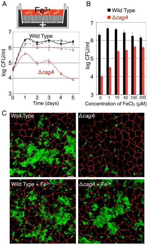
(A) Addition of iron allows ΔcagA growth on the apical cell surface. Polarized cells in the Transwell system were infected with WT or ΔcagA. Co-culture media (+) was added basally. Solid lines indicate conditions with DMEM apically. Dashed lines indicate conditions with 100 µM ferric chloride added to the apical DMEM. Samples were taken daily from the apical chamber and plated for CFU counts. (B) ΔcagA response to iron is concentration dependent. Polarized cells in the Transwell system were infected with WT or ΔcagA. Co-culture media was added basally, and different amounts of ferric chloride (FeCl3) in DMEM added to the apical chamber. Graph shows Hp CFU counts from the apical chamber at day 5 post-infection. (C) Exogenous iron rescues microcolony growth of ΔcagA on the cell surface. 3D confocal images of WT or ΔcagA colonizing the polarized epithelium 5 days post-infection, in the absence or presence of 100 µM ferric chloride (Fe3+). Bacteria are visualized with anti-Hp antibodies (green) and cell junctions are stained red (anti-ZO-1). Scale bar 10 µm. The rescue of ΔcagA growing on the cell surface by iron suggests that CagA affects host epithelial cell function to allow Hp access to micronutrients that are found in the epithelium or across its barrier.
Hp utilizes the polarized epithelium as a filter for the extraction of iron
Host epithelial cells acquire iron largely via transferrin receptor-mediated endocytosis [22]. In the serum where transferrin is normally found, the population of transferrin molecules is 20% – 40% saturated with iron [23], [24]. We thus wondered whether Hp growing on the cell surface gain access to interstitial transferrin and utilize this form of iron for survival. However, in vitro reports have shown that Hp cannot grow on partially saturated transferrin [21]. We found that Hp growth on the cell surface was inhibited by the presence of partially saturated transferrin in the apical medium, indicating that Hp is unable to compete with partially saturated transferrin for iron even in the presence of cells (Figure 2A).
Fig. 2. Holotransferrin promotes Hp microcolony growth on the cell surface. 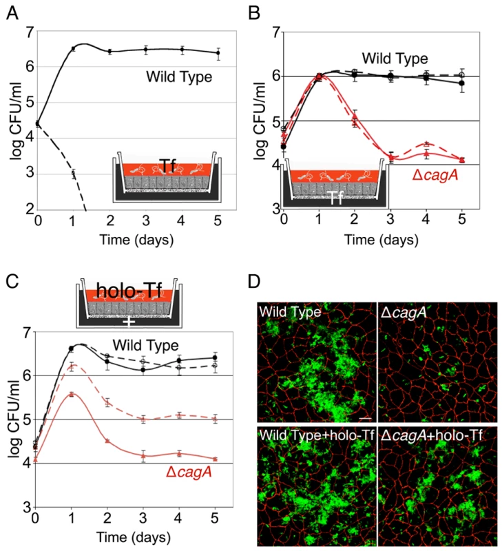
(A) Partially saturated transferrin inhibits Hp colonization of the apical cell surface. Polarized cells in the Transwell system were infected with WT. Co-culture media was added basally. Solid line indicates conditions where DMEM was present apically. Dashed line represents conditions where 75 µg/ml partially saturated transferrin (Tf) was added to the apical DMEM. Samples were taken daily from the apical chamber and plated for CFU counts. (B) Apical Hp microcolonies are protected by the epithelium from partially saturated transferrin in the basal chamber. Polarized cells in the Transwell system were infected with WT or ΔcagA. Solid lines indicate conditions where DMEM was present apically and co-culture media basally. Dashed lines represent conditions where DMEM was present apically and 75 µg/ml partially saturated transferrin (Tf) added to the co-culture media basally. Samples were taken and plated as in (A). (C) Holotransferrin partially rescues ΔcagA growth on the apical cell surface. Polarized cells in the Transwell system were infected with WT or ΔcagA. Co-culture media (+) was added basally. Solid lines indicate conditions with DMEM apically. Dashed lines indicate conditions with 75 µg/ml holotransferrin (holo-Tf) added to the apical DMEM. Samples were taken and plated as in (A). (D) ΔcagA forms microcolonies on the apical cell surface in the presence of holotransferrin. 3D confocal images of WT or ΔcagA colonizing the polarized epithelium 5 days post-infection, in the absence or presence of 75 µg/ml holotransferrin (holo-Tf) in the apical media. Bacteria are visualized with anti-Hp antibodies (green) and cell junctions are stained red (anti-ZO-1). Scale bar 10 µm. Since Hp is susceptible to iron chelation by transferrin, and because transferrin is the major chelator of iron in extracellular fluids of the interstitial space, these results suggest that CagA's mechanism of action is not simply to injure the epithelium and provide paracellular diffusion of transferrin. We wondered if, instead, Hp utilizes the epithelial barrier as a shield from noxious macromolecules in the interstitial space, and whether the bacteria can actively obtain nutrients from the epithelium. To test this, we added transferrin to the basolateral co-culture medium at a concentration sufficient to chelate serum iron and inhibit Hp growth in broth (Figure S1B), and determined if the basal transferrin would inhibit growth of the apical bacteria. As shown in Figure 2B, transferrin at a concentration that quickly kills Hp in the apical chamber had no inhibitory effect across the polarized epithelium, indicating that Hp interaction with the apical cell surface allows the bacteria to utilize the epithelium as a filter for iron.
Epithelial cells selectively acquire transferrin saturated with iron (holotransferrin) via endocytosis, as its affinity for the transferrin receptor is 2000X greater than iron-free transferrin [22]. Since Hp growing on the cell surface are protected from the toxic effects of partially saturated transferrin by the epithelial barrier, we wondered whether the bacteria can obtain iron from holotransferrin, the form of transferrin taken up into cells. A recent report suggested that, in a chemically defined in vitro media system, Hp can obtain iron from holotransferrin, but not partially saturated transferrin [21]. We confirmed these results and tested whether holotransferrin affected the growth of Hp microcolonies on the cell surface by adding it to the apical medium of the Transwell culture system. Not only was holotransferrin not toxic to WT microcolonies, addition of holotransferrin to the apical chamber in fact led to partial rescue of ΔcagA (Figures 2C and 2D). These experiments indicate that Hp growing on the cell surface are able to utilize iron from holotransferrin, even though they cannot compete with partially saturated transferrin for iron, and suggest the hypothesis that acquisition of holotransferrin from within host cells is one mechanism by which Hp acquire iron during mucosal colonization.
Hp colonization of the apical cell surface increases internalized transferrin in a CagA-dependent manner
Since CagA has multiple effects on epithelial physiology and appears to aid Hp in iron acquisition from host cells, we asked if Hp colonization of the apical cell surface affects host cell transferrin recycling. To study this, we utilized MDCK cells stably expressing human transferrin receptor [25], as this allowed us to visualize and quantify transferrin binding to its receptor and its uptake through the use of human transferrin conjugated to a fluorophore, which is readily available (Figure S2A). These cells stably expressing human transferrin receptor formed polarized monolayers, and WT Hp colonized the apical cell surface while ΔcagA exhibited a 100X defect in colonization ability, as seen with untransfected MDCK cells (Figure S2B).
Polarized MDCK cells stably expressing human transferrin receptor were either left uninfected or infected with WT or ΔcagA for two days before fluorescent transferrin assays were performed. In this assay, internalization of transferrin was synchronized by first adding transferrin to the basal chamber on ice. This allowed basolateral binding of transferrin to its receptor, but inhibited its uptake. Unbound transferrin was subsequently washed away, and the cells were warmed to 37°C to allow endocytosis to proceed. As expected, endocytosis was inhibited on ice and transferrin bound to the basolateral membranes of the polarized cells (Figure 3A, top panels) [22], [25]. There was no significant difference in the amount of transferrin bound in WT vs. ΔcagA-infected monolayers (Figure 3A, top panels). Also, by immunoblot, the expression level of the transferrin receptor under the different conditions was similar (Figure 3B). However, when endocytosis was allowed to proceed at 37°C for 30 minutes, significantly higher amounts of transferrin were observed inside the WT-infected monolayers as compared to uninfected or ΔcagA-infected monolayers (Figures 3A and 3C). We found similar results in Caco-2 cells (human colon carcinoma cells) (Figure S3A), indicating that these results are recapitulated in multiple epithelial lines. To determine if this difference is due to CagA vs. the density of colonizing bacteria on the cell surface, we repeated this experiment while adding exogenous iron to the apical chamber to rescue the growth defect of ΔcagA. In these conditions, the numbers of WT and ΔcagA growing on the cell surface were similar, yet the same difference in internalized transferrin was observed (Figures S3B and S3C). Finally, complementation of ΔcagA with the cagA gene (CagA*) led to restoration of the phenotype of increased transferrin internalization on infection with the bacteria (Figure 3C). These experiments suggest that CagA delivery into host cells increases the amount of internalized transferrin.
Fig. 3. Hp colonization of the apical cell surface increases internalized transferrin. 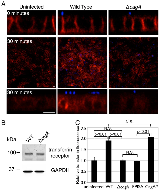
(A) 3D confocal images of fluorescent transferrin signal in polarized MDCK cells stably expressing human transferrin receptor, uninfected or infected with WT or ΔcagA. Fluorescent transferrin was added to the basal chamber and incubated on ice for 30 minutes, unbound transferrin washed away, then immediately fixed (top panels, cross-sectional view, 0 minutes post-uptake), or further incubated for 30 minutes at 37°C to allow uptake of bound transferrin (middle and bottom panels, top and cross-sectional views, 30 minutes post-uptake). Fluorescent transferrin is shown in red, and bacteria are visualized with anti-Hp antibodies (blue). Scale bars 10 µm. (B) Hp colonization does not significantly affect host cell transferrin receptor expression. Polarized cells in the Transwell system were infected for 2 days with WT or ΔcagA. Whole-cell lysates from these infections were separated by SDS-PAGE, transferred to a nitrocellulose membrane, then immunoblotted with antibodies against transferrin receptor (top panel) and against GAPDH as a loading control (bottom panel). (C) Quantification of transferrin fluorescence 30 minutes post-uptake in polarized epithelial monolayers. Monolayers were infected for 2 days with the indicated Hp strains, fixed after 30 minutes of transferrin uptake, and total transferrin fluorescence measured from multiple 3D confocal images. EPISA is a mutant expressing a mutated CagA that cannot be phosphorylated. CagA* is the complemented ΔcagA mutant. p-values were obtained with a Mann-Whitney statistical test. N.S. indicates no statistical significance. Once injected into the host cell, CagA is tyrosine phosphorylated by host Src - and Abl-family tyrosine kinases at several repeated sites in the C-terminal end containing EPIYA motifs [26]–[28]. CagA then acts as an adaptor protein that stimulates signaling downstream of growth factor receptor tyrosine kinases [29]–[32]. Since growth factor signaling increases transferrin uptake [33], we studied whether CagA phosphorylation is necessary for its effects on transferrin internalization. We found that the ability of CagA to increase internalized transferrin depends on the presence of the EPIYA motifs, as infection of polarized monolayers with a mutant lacking these phosphorylation domains resembled ΔcagA infection (Figure 3C).
These results indicate that CagA injected by Hp microcolonies on the apical cell surface increases transferrin internalization through receptor tyrosine kinase-like signaling, and suggest that this leads to an increased availability of iron for the colonizing bacteria. However, under normal conditions, transferrin should not be released to the apical side of an epithelium, since its recycling is confined to the basolateral membrane where the receptor is exclusively found [22]. This suggests that infecting bacteria perturb not just uptake, but also localization of the transferrin/transferrin receptor complex.
Transferrin receptor is mislocalized to sites of Hp microcolony growth at the apical cell surface
Hp is known to affect host cell polarity and intracellular trafficking [34]–[38], and our previous study showed that perturbation of host cell polarity is involved in enhancing colonization of the polarized epithelium [8]. We therefore wondered if Hp colonization might lead to mis-sorting of the transferrin receptor, and hence transferrin and possibly iron, to sites of bacterial microcolony growth on the apical surface.
To address this, we fixed uninfected or infected polarized MDCK monolayers in conditions that do not permeabilize the membrane and then applied antibodies against the transferrin receptor only to the apical side. In this manner, we can detect whether small amounts of transferrin receptor are found at the apical membrane without detecting internalized or basolateral transferrin receptor. We observed distinct puncta of immunolabeled transferrin receptor localized at the apical membrane near the Hp microcolonies (Figure 4A). This mislocalization of the transferrin receptor does not occur immediately after bacterial attachment to the apical surface, since monolayers fixed after 5 minutes of infection did not show puncta of transferrin receptor on the apical side (Figure S4A). This implies that Hp can affect host cell polarity locally to mislocalize basolateral proteins to sites of microcolony growth.
Fig. 4. Transferrin receptor is mislocalized apically to sites of bacterial microcolonies. 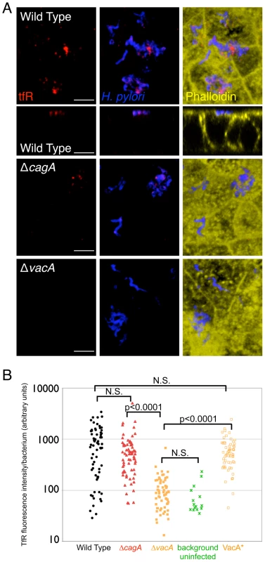
(A) Polarized MDCK cells in the Transwell system were infected with WT, ΔcagA, or ΔvacA for 2 days. Apical staining with anti-transferrin receptor antibodies was carried out on non-permeabilized samples. Bacteria are visualized with anti-Hp antibodies (blue), transferrin receptor (tfR) is stained red, and phalloidin staining of f-actin is shown in yellow. 3D confocal images are shown, and cross-sectional view is also presented for WT (second row). Scale bars 5 µm. (B) Quantitative data of the transferrin receptor (tfR) fluorescence intensity associated with the bacterial microcolonies, determined by the fluorescence voxel volume of each microcolony stained with anti-Hp antibodies, and the fluorescence sum of the transferrin receptor signal associated with the microcolonies, measured from multiple 3D confocal images. VacA* is the complemented ΔvacA mutant. Each point on the graph represents a microcolony. p-values were obtained with a Mann-Whitney statistical test. N.S. indicates no statistical significance. To confirm these findings, we used a different technique that allows selective biotinylation of surface proteins of the polarized epithelium (Figure 5A) [39], [40]. We selectively biotinylated the basolateral surface on ice, and then allowed internalization and recycling of the biotinylated basolateral proteins for 30 minutes at 37°C. We then stained the apical surface without permeabilization with fluorophore-conjugated streptavidin to detect mislocalized basolateral proteins. As with the previous results, we found that Hp microcolonies are associated with membrane patches containing basolateral proteins that were mis-sorted to the apical membrane (Figures 5B and 5C, bottom panels, and Figure 5D). To confirm that endocytosis is required, we repeated the staining on infected monolayers that were fixed immediately after surface biotinylation on ice. These monolayers did not contain apical biotin staining (Figures 5B and 5C, top panels, and Figure 5D). To determine whether this phenomenon is generalizable to other polarized cell models, we repeated the staining of transferrin receptor in Caco-2 cells. We obtained similar results as with MDCK cells, indicating that Hp can induce mis-sorting of the transferrin receptor in multiple epithelial lines (Figure S5). Finally, to determine whether mislocalization of basolateral proteins is restricted to a subset of proteins or affects all basolateral proteins, we used antibodies to other basolateral markers. E-cadherin is a cell-cell adhesion molecule that is normally absent from the apical membrane. Using apical staining of non-permeabilized cell monolayers with E-cadherin antibodies, we had previously shown that E-cadherin is absent from most of the apical membrane except for sites of cell extrusion [41]. When we applied these antibodies to Hp-infected monolayers, we did not find E-cadherin associated with Hp microcolonies (Figure S4B), indicating that not all basolateral proteins are mislocalized during Hp colonization.
Fig. 5. Hp induces mislocalization of basolateral proteins to the apical surface at sites of bacterial attachment. 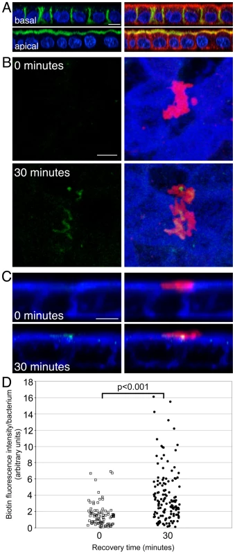
(A) Cell surface proteins of polarized cells on Transwell filters were selectively labeled with biotin in either the basal (top panels) or apical (bottom panels) chambers. Streptavidin labels biotinylated proteins in green, phalloidin stains f-actin in red and DAPI stains cell nuclei in blue. Scale bar 5 µm. (B and C) Polarized cells infected apically with WT for 2 days were selectively biotinylated on ice at the basolateral surface, and either immediately fixed (top panels, B and C), or first incubated for 30 minutes at 37°C (bottom panels, B and C) before fixation and apical streptavidin staining. Top views (B) and cross-sectional views (C) are shown. Bacteria are visualized with anti-Hp antibodies (red) and biotin is marked in green. Phalloidin staining of f-actin is shown in blue. Scale bars 5 µm. (D) Quantification of basolateral proteins associated with Hp microcolonies. Open squares are data from monolayers fixed immediately after basolateral biotinylation. Closed circles are data from monolayers incubated at 37°C for 30 minutes after basolateral biotinylation. Each point represents the total fluorescence intensity of apically-exposed biotin associated with a microcolony, divided by the number of bacteria present in that microcolony. p-value was obtained with a Mann-Whitney statistical test. Examination of monolayers infected with ΔcagA indicated that CagA-deficient bacteria are less efficient but still able to recruit transferrin receptor to the sites of bacterial microcolonies (Figure 4). Quantification of the amount of transferrin receptor observed apically at the sites of ΔcagA microcolonies showed that the median amount (514 arbitrary units/bacterium) was less than that observed with WT (774 arbitrary units/bacterium), but this was not statistically significant (Figure 4B).
The ability of ΔcagA to still cause mislocalization of host cell transferrin receptor apically to sites of bacterial microcolonies suggested that other bacterial factor(s) may be involved in this phenomenon. Another major virulence factor of Hp, vacuolating cytotoxin (VacA), has been reported to interfere with the endocytic pathway of host cells [38]. We therefore decided to test the role of VacA in the mislocalization of transferrin receptor. We constructed a VacA-deficient Hp mutant (ΔvacA), and found that in monolayers infected with ΔvacA, transferrin receptor was absent from the sites of bacterial microcolonies on the apical cell surface (Figure 4). This result was confirmed in infected Caco-2 cell polarized monolayers (Figure S5). We also complemented ΔvacA with the vacA gene, and found that this reconstitution (VacA*) restored the ability of the bacteria to mislocalize transferrin receptor apically to sites of bacterial microcolonies (Figure 4B).
Selective biotinylation of basolateral epithelial proteins and quantification of the amount of biotin subsequently associated with apical microcolonies indicated that both ΔcagA and ΔvacA had significantly less biotin associated with microcolonies than WT (Figure S6). This suggests that both CagA and VacA are involved in disruption of polarity, and that the transferrin receptor is not the only molecule that is mislocalized apically. We also found that a mutant deficient in both CagA and VacA (ΔcagAΔvacA) was still able to mislocalize proteins to the sites of bacterial microcolonies (Figure S6), implying that CagA and VacA are not the only bacterial factors involved in this process, although they do appear to play major roles.
Together, these results indicate that Hp colonization of the apical cell surface leads to mislocalization of a subset of basolateral proteins to the apical cell membrane at sites of bacterial microcolony growth, with the Hp virulence factors CagA and VacA playing major roles in this process. One of the proteins mislocalized to these sites is the transferrin receptor, and its mislocalization is primarily dependent on VacA.
VacA aids Hp colonization of the apical cell surface
Given the role of VacA in transferrin receptor mislocalization, we next tested whether VacA affects the ability of Hp to colonize the apical cell surface of a polarized epithelium. We observed that ΔvacA have approximately a 10X decrease in bacterial counts at day 5 post-infection, as compared to WT (Figure 6A). In the presence of rich media in the apical chamber, ΔvacA grow as well as WT, indicating that this phenotype is not due to an in vitro growth defect of the mutant (Figure S7).
Fig. 6. VacA contributes to Hp colonization of the apical cell surface. 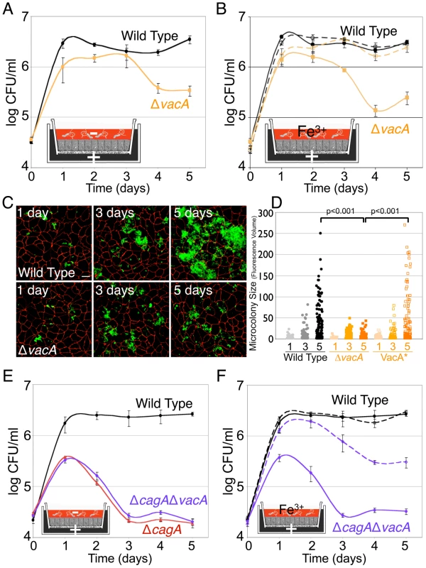
(A) VacA aids Hp colonization of the apical cell surface. Using the Transwell system, cells were infected with WT or ΔvacA, and co-culture media added only to the basal chamber (+). DMEM was added to the apical chamber (−). Samples were taken daily from the apical chamber and plated for CFU counts. (B) Exogenous addition of iron apically rescues ΔvacA growth on the polarized epithelium. Polarized cells were infected as in (A). Solid lines indicate conditions with DMEM apically. Dashed lines indicate conditions with 100 µM ferric chloride (Fe3+) added to the apical DMEM. Samples were taken and plated as in (A). (C) 3D confocal images of WT or ΔvacA colonizing the cell surface of polarized MDCK cells in the Transwell system. Cells were infected for 5 minutes and then unattached bacteria washed away and media replaced. At 1, 3 and 5 days post-infection, samples were fixed and processed for immunofluorescence. Bacteria are visualized with anti-Hp antibodies (green) and cell junctions are stained red (anti-ZO-1). Scale bar 10 µm. (D) Quantification of WT, ΔvacA and VacA* microcolony sizes over time (1, 3 and 5 days), determined by fluorescence volume measured from multiple 3D confocal images. VacA* is the complemented ΔvacA mutant. Each point on the graph represents a microcolony. p-value was obtained with a Mann-Whitney statistical test. (E) CagA and VacA work in concert to enable Hp colonization of the polarized epithelium. Polarized cells were infected as in (A) with the strains indicated. Samples were taken and plated as in (A). (F) CagA and VacA work together to aid Hp acquisition of iron from host cells. Polarized cells were infected as in (A). DMEM (solid lines) or DMEM + 100 µM ferric chloride (Fe3+, dashed lines) was added apically. Samples were taken and plated as in (A). To determine whether the decrease in bacterial counts correlates with a defect of colonization of the apical cell surface, we examined microcolony formation on polarized cells infected with ΔvacA by confocal immunofluorescence microscopy. Initial adherence of ΔvacA to the apical cell surface was no different from WT, with an average of 9 bacteria adhered/100 cells in each case (p = 0.5). However, ΔvacA formed significantly smaller microcolonies on the cell surface at day 5 post-infection (Figures 6C and 6D). Complementation of ΔvacA (VacA*) led to restoration of the ability of the bacteria to effectively colonize the polarized epithelium, with the formation of large microcolonies (Figure 6D).
We also tested whether the ΔvacA mutant is defective in iron acquisition from the host, by supplementing the apical media with iron. Exogenous addition of iron to the apical chamber led to rescue of the colonization defect shown by ΔvacA (Figure 6B). To examine if CagA and VacA act in similar or different pathways in aiding Hp in cell surface colonization, we tested the mutant deficient in both CagA and VacA (ΔcagAΔvacA). This double mutant resembled the single CagA-deficient mutant, and exogenous addition of iron apically partially rescued its ability to colonize the polarized epithelium (Figures 6E and 6F). The phenotype observed is not due to the mutant having an in vitro growth defect, as growth of this double mutant was similar to WT in the presence of rich media added to the apical chamber (Figure S7).
These results indicate a role for VacA in Hp colonization of the polarized epithelium, and suggest that CagA and VacA work in concert to aid Hp acquisition of iron from, and colonization of, host polarized epithelial cells.
Host cell transferrin receptor is involved in Hp microcolony formation on the apical cell surface
The data presented above indicate that the Hp virulence factors CagA and VacA both affect normal recycling and trafficking of the transferrin/transferrin receptor complex, which is the major iron uptake mechanism of epithelial cells [22]. Furthermore, mutants in both virulence factors are defective in colonizing the polarized cell surface and these defects can be partially rescued by exogenous addition of iron. This suggests that Hp perturbation of the transferrin/transferrin receptor recycling pathway might be used by the bacteria to obtain iron from host cells.
To directly test if the transferrin receptor pathway is important for Hp colonization of the polarized epithelium, we silenced expression of the transferrin receptor during infection and asked whether this affects microcolony growth. We designed siRNAs directed against the canine transferrin receptor and selected two that produce very effective knockdown of transferrin receptor expression (Figure 7A). Cells transfected with a mixture of these two siRNAs were seeded on Transwell filters and allowed to polarize before infection with WT. A first observation was that Hp was able to recruit the minimal amount of transferrin receptor still expressed by the host cells to the sites of bacterial microcolonies (Figure 7B). More importantly, Hp formed significantly smaller microcolonies on the apical cell surface of monolayers where transferrin receptor expression had been knocked down, as compared to monolayers transfected with a control siRNA (eGFP) (Figures 7C and 7D).
Fig. 7. Down-regulation of host cell transferrin receptor decreases Hp microcolony growth on the cell surface. 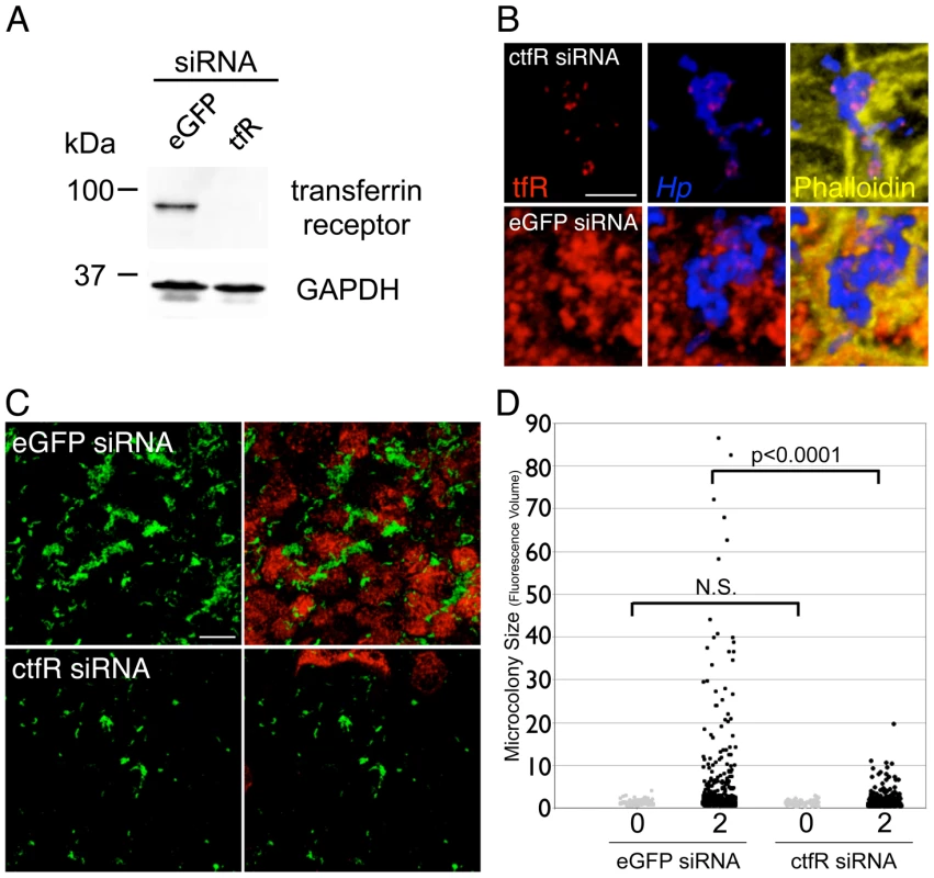
(A) siRNA knockdown of transferrin receptor expression in MDCK cells. Cells were transfected with a combination of two siRNAs directed against transferrin receptor, or with siRNA directed against enhanced GFP (eGFP) as a control. 3 days post-transfection, the cells were collected and lysates separated by SDS-PAGE, transferred to a nitrocellulose membrane, then immunoblotted with antibodies against transferrin receptor (top panel) and against GAPDH as a loading control (bottom panel). (B) Residual transferrin receptor is mislocalized apically to sites of bacterial microcolonies. MDCK cells were transfected with siRNA against canine transferrin receptor (ctfR, top panels) or eGFP as a control (bottom panels). After polarization, the cells were infected with WT for 1 day. Bacteria are visualized with anti-Hp antibodies (blue), transferrin receptor (tfR) is stained red, and phalloidin staining of f-actin is shown in yellow. Scale bar 5 µm. (C) Hp form smaller microcolonies on the apical cell surface when transferrin receptor expression is knocked down. 3D confocal images of WT colonizing the polarized epithelium 2 days post-infection, on cells either transfected with siRNAs against canine transferrin receptor (ctfR) or eGFP as a control. Bacteria are visualized with anti-Hp antibodies (green) and transferrin receptor is stained red. Scale bar 10 µm. (D) Quantification of Hp microcolony sizes on cells transfected with siRNAs directed against transferrin receptor or eGFP as a control. Data from 0 and 2 days post-infection are shown. Microcolony sizes were determined by fluorescence volume measured from multiple 3D confocal images. Each point on the graph represents a microcolony. p-values were obtained with a Mann-Whitney statistical test. N.S. indicates no statistical significance. We made use of the fact that the siRNAs directed against the canine transferrin receptor are highly specific and do not cross-react with human transferrin receptor (Figure S8A) to test that the phenotype observed was not due to off-target effects of the siRNAs. In MDCK cells stably expressing human transferrin receptor, knockdown of endogenous canine transferrin receptor expression left expression of human transferrin receptor intact (Figure S8A). Hp allowed to colonize the apical surface of these cells formed microcolonies similar in size to those formed by Hp colonizing cells transfected with a control siRNA (Figure S8B). This indicates that the decreased ability of Hp to colonize the apical cell surface after knockdown of transferrin receptor expression in MDCK cells is specifically due to decreased expression of transferrin receptor in those cells.
Collectively, these results show that host cell transferrin receptor is functionally important in enabling Hp colonization of the apical surface of a polarized epithelium. They also suggest that CagA and VacA-mediated perturbation of transferrin/transferrin receptor recycling allows Hp to acquire iron from the host cells.
Hp colonization of the polarized epithelium leads to increased apical release of transferrin
Our data suggest a model in which Hp colonization of the apical cell surface leads to mis-sorting of a subset of the transferrin/transferrin receptor complex and transcytosis of the complex from the basolateral to the apical surface at the sites of bacterial microcolony growth. We determined whether transferrin is transcytosed apically by adding biotinylated transferrin to the basolateral media and then assaying for the presence of biotinylated transferrin in the apical chamber after a 24-hour incubation period. We detected a 1.5-fold increase in the amount of biotinylated transferrin in the apical chamber of WT-infected monolayers as compared to uninfected monolayers (Figure 8 and Figure S9). This increase was dependent on CagA, since monolayers infected with ΔcagA had similar amounts of biotinylated transferrin in the apical chamber as uninfected monolayers (Figure 8). To determine that this increase was due to transcytosis, and to control for possible paracellular leakage of macromolecules, we also added biotinylated albumin to the basolateral chamber. In contrast to transferrin, the small amount of biotinylated albumin detected in the apical chamber was the same irrespective of whether the monolayers were infected with WT or ΔcagA, or left uninfected (Figure 8 and Figure S9). Infection with the complemented ΔcagA (CagA*) resulted in restoration of the phenotype of increased transferrin transcytosis (Figure 8).
Fig. 8. Hp colonization of the cell surface leads to transferrin transcytosis. 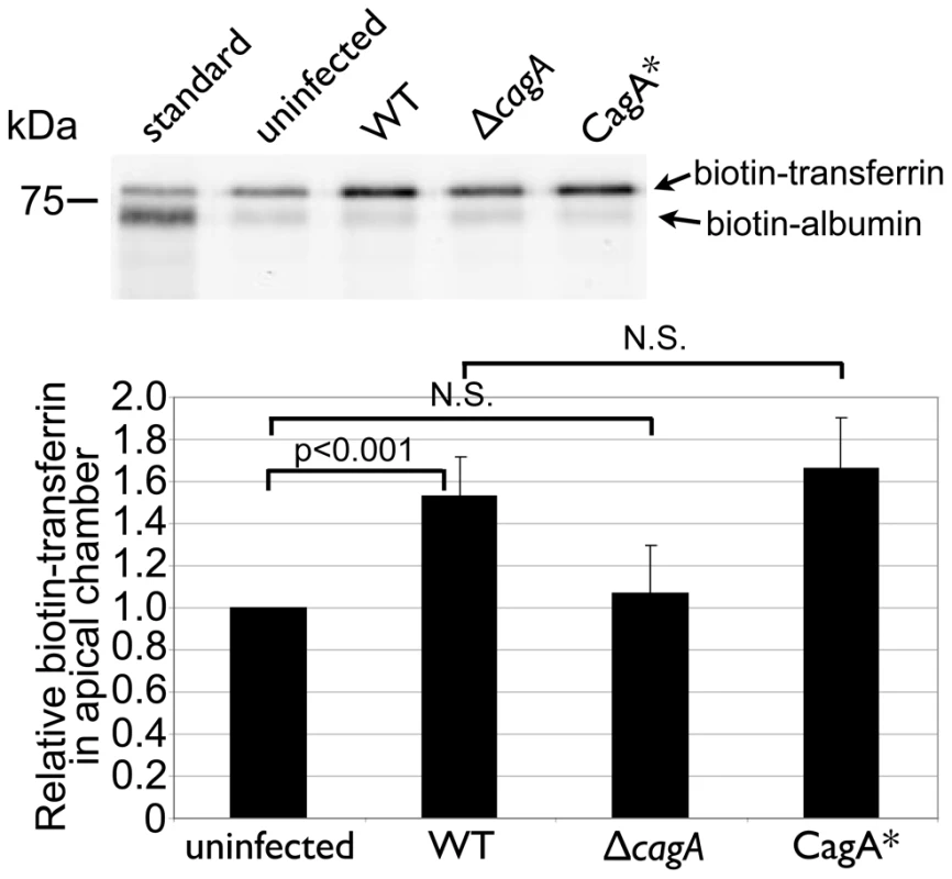
30 µg of biotin-albumin and 75 µg of biotin-transferrin were added to the basal chamber of uninfected or 2 day-infected polarized monolayers. The apical supernatant was sampled after a 24 hour incubation, and 10 µl of these samples separated by SDS-PAGE, blotted onto nitrocellulose, and the biotinylated albumin and transferrin were visualized with fluorescent streptavidin. The lane labeled “standard” is a 1∶250 dilution of the basal media containing biotin-albumin + biotin-transferrin. CagA* is the complemented ΔcagA mutant. Each band was quantified with the LI-COR Odyssey Scanner. Biotin-albumin amounts were used to normalize for loading. The graph depicts the average result from 6 experiments. p-values were obtained with a Wilcoxon signed rank test, using a hypothetical median of 1. N.S. indicates no statistical significance. These findings indicate that Hp colonization of the apical cell surface results in transcytosis of transferrin to the apical side of the cell.
CagA confers an adaptive advantage in colonization of the stomach under iron-deplete conditions
Hp colonizes multiple niches in the stomach (i.e. free-swimming in the mucus layer vs. the cell surface), and since CagA-negative strains are common in nature, it is likely that in vivo, Hp utilizes multiple modes of iron acquisition. However, our findings suggest that CagA may be more important for the bacteria when colonizing hosts that are iron-depleted or during conditions of poor dietary iron content. To address this question, we utilized the Mongolian gerbil model of Hp infection to determine if the iron status of the host would affect WT or ΔcagA colonization.
Mongolian gerbils were maintained either on a regular diet containing 250 ppm iron, or an iron-deficient diet containing <1 ppm iron, for 3 weeks prior to infection, and then through the duration of the infection (see Materials and Methods for details). Dietary restriction of iron has been shown to result in decreased host iron levels in mice [42]–[44], and we verified that the animals placed on the iron-deficient diet had reduced iron levels, as measured by inductively coupled plasma-mass spectrometry of liver samples (Figure S10A). For these infections, we used the Hp strain 7.13, which has been previously shown to reproducibly colonize the Mongolian gerbil stomach, is able to deliver CagA into host cells, and whose isogenic ΔcagA mutant exhibits a defect in colonization of the polarized epithelium in vitro, similar to the Hp strain G27-MA [8], [45]. In conditions of growth in nutrient-rich broth, 7.13 WT and ΔcagA grow with similar kinetics (Figure S10B). We infected Mongolian gerbils with either 7.13 WT or ΔcagA, and examined bacterial loads at 6 or 8 weeks after infection. 7.13 WT colonized iron-replete and iron-deficient animals to similar levels (Figure 9). The isogenic ΔcagA mutant also colonized iron-replete animals to a similar level as WT. However, 7.13 ΔcagA showed a significant decrease in colonization levels in the stomachs of iron-deficient animals, both in comparison to WT in iron-deficient animals and in comparison to ΔcagA in iron-replete animals (Figure 9). We also found that bacteria could not be recovered from 9/16 animals in the ΔcagA-infected iron-deficient group of animals, unlike the WT-infected iron-deficient group, in which Hp could be recovered from all 14 animals infected, at the end of the experiment (Figure 9).
Fig. 9. Host iron depletion decreases ΔcagA fitness in vivo. 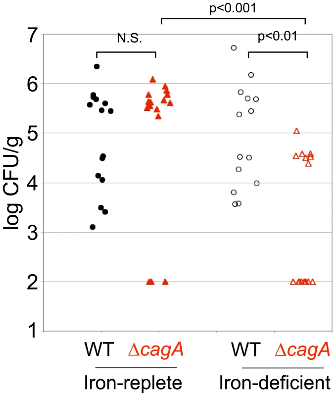
Mongolian gerbils maintained either on a regular, iron-replete diet, or on an iron-deficient diet were infected with Hp strain 7.13 WT or its isogenic ΔcagA mutant. 6–8 weeks post-inoculation, the animals were sacrificed and bacterial counts obtained from the stomach. Each point on the graph represents one animal. Animals from which no bacteria could be recovered are represented at 100 CFU/g, which is the limit of detection. p-values were obtained with a Mann-Whitney statistical test. N.S. indicates no statistical significance. These results mirror our in vitro findings that CagA plays an important role in enabling effective acquisition of iron from the host during Hp colonization of its gastric epithelial niche.
Discussion
Bacterial virulence factors are defined as molecules associated with disease. The major virulence factors of Hp, CagA and VacA, are epidemiologically linked to disease and possess multiple biological properties that can be deleterious to host cells [46]–[49]. However, understanding how these molecules function during an infection requires asking not just how virulence factors disrupt the host cell, but also how such effects benefit the bacteria. Our previous study had established that Hp can grow as microcolonies attached to the apical cell surface of a polarized epithelium, even in conditions where the free-swimming bacteria are rapidly killed [8]. CagA helps the bacteria form microcolonies and exploit this niche by perturbing cell polarity [8]. Here, we extend the concept that bacteria perturb host cell polarity to use the apical cell surface as a replicative niche. We show that bacterial virulence factors alter polarized host intracellular trafficking, suggesting a novel mechanism by which the bacteria are able to acquire essential micronutrients from host cells and colonize the apical cell surface.
We found that exogenous iron added to the media bathing bacterial microcolonies on the apical cell surface partially rescues the Hp ΔcagA mutant growth defect. This suggests that one of the factors Hp extract from epithelial cells is iron, and that one of CagA's benefits to the bacteria involves facilitating iron acquisition from the epithelium. Furthermore, Hp is able to colonize the apical membrane of the epithelium even in the presence of excess iron chelators bathing the interstitial side of the epithelium, suggesting that it can acquire iron directly from the host cells without gross disruption of epithelial integrity. We note that attachment of Hp to non-polarized host cells had previously been reported to result in upregulation of expression of several annotated iron uptake proteins [50]. Because uptake of iron into host epithelial cells is a polarized process, occurring largely basolaterally through the transferrin/transferrin receptor recycling pathway [22], we probed whether this pathway is manipulated by Hp during colonization of the cell surface.
In the presence of an intact epithelial barrier, microbes on the apical surface do not have access to interstitial iron-bound transferrin or its recycling. In addition, partially saturated transferrin, which is the form found in the interstitium, is toxic to Hp [21]. However, Hp is able to utilize iron from holotransferrin [21], which is the form that eukaryotic cells preferentially bind and uptake due to its higher affinity for the transferrin receptor, as compared to partially saturated or iron-free transferrin [22], [51]. This suggested that Hp may be able to utilize the epithelium both as a barrier against the toxic effects of partially saturated transferrin, and as a source of holotransferrin. We observed that the apical cell surface colonization defect of ΔcagA mutants can be partially rescued by addition of holotransferrin to the apical chamber, suggesting that intracellular holotransferrin could be one possible iron source for colonizing bacteria.
For Hp microcolonies to utilize holotransferrin as an iron source without destroying the epithelium, polarized uptake and recycling of transferrin would have to be perturbed. Both CagA and VacA have biological properties that could be involved in this process. For example, CagA is known to be able to induce receptor tyrosine kinase (RTK)-like signaling [29]–[31], and growth factor RTK activation has been shown to increase uptake of transferrin in other models [33]. CagA has also been shown to affect cell polarity and the assembly of the epithelial junctions [35]–[37], [45], both of which could influence sorting of basolateral molecules [52]–[54]. VacA, another major virulence factor of Hp, has previously been reported to affect endosomal trafficking [38]. We therefore hypothesized that CagA and VacA could have effects on the host cell transferrin/transferrin receptor recycling pathway.
We found that CagA injected into host cells by Hp growing as microcolonies on the apical cell surface increased internalized transferrin. This required signaling via the EPIYA motifs in the C-terminus of the protein, which are important in the activation of RTK-like signaling [29], [30], [32]. We also observed that a subset of the transferrin receptor population is mislocalized from the basolateral surface specifically to sites of bacterial microcolonies on the apical cell surface. In our model system, this mislocalization is largely dependent on the action of VacA, as a ΔvacA Hp mutant failed to recruit transferrin receptor apically. We were able to show a direct involvement of the transferrin/transferrin receptor recycling process in the colonization of the polarized epithelium by Hp, since siRNA knockdown of transferrin receptor expression resulted in decreased growth of Hp microcolonies on the cell surface. Our model suggests that Hp colonizing the apical cell surface induce mis-sorting of a subset of the transferrin/transferrin receptor complex apically. In accord with this, we observed significantly increased transcytosis of transferrin from the basal to the apical compartment and its release into the apical media when the epithelium is infected by WT Hp.
Several important questions remain regarding this model. First, it is clear that iron acquisition from the epithelium is only one of several mechanisms of iron acquisition by Hp. Hp can live as a free-swimming population in vitro without the need for contact with epithelial cells. In vivo, Hp may obtain iron through several mechanisms and from various sources. For example, Hp residing as free-swimming bacteria in the mucus layer may obtain iron conjugated in mucus glycoproteins, or perhaps from the release of dietary iron by the acidic stomach lumen, or from inflammatory exudates, although these potential sources need study. Of note is that in addition to its role in causing peptic ulcers and its association with gastric cancer, Hp infection has recently been associated with iron deficiency anemia unrelated to blood loss [3]. Indeed, the latest Maastricht Consensus Report recommends diagnosis and treatment of Hp in cases of unexplained iron deficiency anemia [55]. There are now several reports in the literature showing that Hp eradication improved or cured previously unexplained cases of iron deficiency anemia [4], [56], [57]. Second, it will also be important in future studies to directly visualize the transfer of iron from the host to the bacteria and the precise molecular mechanisms involved. The observation that specific basolateral molecules, such as the transferrin receptor, are highly concentrated at sites of bacterial microcolonies in comparison to the rest of the apical membrane suggests that these sites are a very specialized and enriched microenvironment. However, methods that allow the visualization and quantification of iron in a spatially defined manner at sub-micrometer resolution are still at early stages of development [58], [59]. Technical limitations such as these, as well as difficulties associated with maintaining a polarized monolayer in which the transferrin receptor or other important host molecules have been downregulated over prolonged periods of time, will first have to be overcome before the exact mechanisms by which Hp extracts iron from the host epithelium can be fully understood. Several of the known effects of CagA and VacA on host cell physiology may have important roles in altering trafficking of transferrin and other molecules for the benefit of colonizing bacteria. For example, we showed here that signaling through the EPIYA motifs of CagA increase host internalization of transferrin. CagA also has multiple effects mimicking growth factor activity which could influence the way in which gastric epithelial cells uptake and process nutrients that could be used by colonizing Hp [29]–[31], [60]. In addition, we had previously reported that CagA's perturbation of host cell polarity via its actions on Par1b are important in enabling Hp colonization of the apical cell surface [8]. Of note, one of Par1b's functions is in organizing microtubules, and it has been speculated to play a crucial role in regulation of intracellular vesicle transport [61]. In addition to its effects on endosomal trafficking, VacA has been shown to act as a pore on membranes, and may independently increase transcellular and paracellular flow of iron and other essential factors [48], [62], [63]. We did not observe transferrin receptor mislocalization to sites of bacterial attachment in preliminary experiments with purified VacA added exogenously to ΔvacA-infected polarized cell monolayers (data not shown). The delivery of VacA can be local through contact by adhered bacteria [64], as well as at a distance through diffusion of soluble VacA [48]. Since localized VacA delivery during cellular infection may not be equivalent to generalized intoxication, it will be interesting to define how these two forms of delivery differ in their effects on polarized epithelia. It is also possible that VacA is necessary, but not sufficient, for this process.
The potential cooperation between CagA and VacA to affect polar transport of micronutrients to colonizing microcolonies is exciting, since CagA and certain VacA alleles have been linked genetically [65], and possible interplay between their functions have been reported [66]–[70]. Different subtypes of CagA and VacA may also have varied activities in aiding Hp colonization of the epithelium. For example, the “East Asian” form of CagA has been reported to bind more strongly to Par1b than the “Western” form of CagA [71]. How do such differences translate to the ability of different strains of the bacteria to colonize the epithelium? Our findings suggest that CagA and VacA may act in concert in a novel way of inducing the host epithelium to relinquish essential micronutrients that does not necessitate destroying the epithelium or causing gross leakage of interstitial macromolecules into the lumen. Further studies will reveal the exact pathways that are usurped by CagA and VacA in altering host transferrin trafficking, visualize how iron is delivered to Hp on the cell surface, and perhaps uncover additional iron acquisition methods by Hp.
Our use of a simplified experimental system allowed us to focus on the microenvironment of the apical cell surface as a replicative niche, and to uncover one possible mechanism by which Hp may obtain iron from host epithelial cells. We subsequently found that ΔcagA mutant Hp have a decreased ability to colonize the stomachs of Mongolian gerbils that are iron-deficient whereas WT bacteria do not, indicating that CagA aids Hp in acquiring iron from the host during in vivo bacterial colonization. Our findings also raise the question of how host micronutrient levels impact the pathogenesis of colonizing Hp. For example, might decreased levels of micronutrients in the host lead to increased pathogenicity of the bacteria? We note here that expression of both cagA and vacA have been reported to be upregulated in conditions of iron starvation in vitro [9], [72], and that the ferric uptake regulator (Fur) protein indirectly regulates vacA expression [72], [73]. Previous epidemiological data had suggested an association between iron deficiency and gastric cancer, although this data predates our understanding of the role of Hp infection [74]. It will now be interesting to examine epidemiologically the possible roles of CagA and VacA in the context of iron deficiency in human populations.
We suggest that manipulation of host epithelial polarity is akin to utilizing the epithelium as a filter to acquire essential micronutrients while maintaining a barrier that protects the microbes from innate immune defenses present in the interstitial space. Perturbation of host epithelial polarity by Hp appears to be a specific and subtle process, not due to a generalized loss of epithelial polarity, since biotinylated proteins from the basolateral membrane form patches specifically under apical bacterial microcolonies, and not all basolateral proteins are found in these membrane patches. Although we focused on transferrin receptor mislocalization in this study, the technique of basal biotinylation of host membrane proteins in infected monolayers suggests that there are multiple proteins that are mislocalized to the apical sites of bacterial growth. The identity of these proteins may shed other important insights into the way microbes colonize the epithelial surface. CagA and VacA play major roles in this mislocalization, but other bacterial factors do appear to be involved as well. The ability to manage host epithelial polarity is emerging as an important facet of bacterial-host interactions. For example, Listeria monocytogenes has been shown to take advantage of cell polarity changes for invasion [41], [75], and mislocalization of basolateral proteins to the apical cell surface as a result of bacterial infection has also been described for other pathogens such as enteropathogenic Escherichia coli and Pseudomonas aeruginosa [76], [77]. What are the mechanisms by which specific basolateral molecules are sequestered to the sites of bacterial microcolonies? During P. aeruginosa infection of polarized epithelia, phosphatidylinositol 3, 4, 5-trisphosphate (PIP3), a basolateral membrane lipid important in cellular signaling and polarity, is mislocalized to the sites of bacterial attachment on the apical cell surface [77], [78]. PIP3 is an intriguing candidate in the context of our results with the transferrin receptor, as it has recently been shown to also localize to recycling endosomes, and to be important for the proper sorting of recycling cargo, such as the transferrin receptor [79]. Furthermore, both CagA and VacA have been implicated in the activation of the phosphatidylinositol 3-kinase pathway, which catalyzes production of PIP3 [80], [81]. It will be interesting to test if PIP3 is recruited to sites of Hp microcolonies on the apical cell surface, and whether PIP3 function is involved in Hp's colonization of the polarized epithelium.
In summary, our results show that iron is one important factor that Hp is able to obtain from host cells during colonization of the apical cell surface, and illustrate a way in which CagA and VacA work in concert to aid the bacteria in establishing a replicative niche. We hypothesize that growth on the epithelial surface involves more than just iron acquisition from the host. For example, it has previously been shown that Hp can acquire cholesterol directly from host cells via contact [82]. We speculate that Hp has evolved sophisticated mechanisms to manipulate host cell physiology for its own benefit, and that these features have side effects that result in disease in a subset of the infected population. Instead of destroying the epithelium as may occur in some types of acute bacterial infection, mucosal colonizers like Hp use more subtle mechanisms of local epithelial perturbation. We propose that mucosal colonizers may be utilizing the polarized epithelium as a “filter” both to protect themselves from potentially toxic host defense molecules, and to selectively extract micronutrients that are present inside the host in a form that is usable by the bacteria. Future exploration of the nature of host proteins associated with apical bacterial microcolonies, and the possible role of other bacterial factors in perturbing polarity, will give us a better understanding of how Hp and other mucosal colonizers affect the epithelial surface for their benefit.
Materials and Methods
Ethics statement
All animal experiments were performed in accordance to NIH guidelines, the Animal Welfare Act, and US federal law. Such experiments were carried out with the approval of the Institutional Animal Care and Use Committee of Vanderbilt University, which has been accredited by the Association of Assessment and Accreditation of Laboratory Animal Care International (AAALAC). All animals were housed in an AAALAC-accredited research animal facility fully staffed with trained technical, husbandry, and veterinary personnel.
Cell culture
Madin-Darby Canine Kidney II (MDCK) cells (kindly provided by W. James Nelson, Stanford University, Stanford, CA) [83], and MDCK cells stably expressing human transferrin receptor (kindly provided by Suhaila White and Suzanne Simon, The Salk Institute, La Jolla, CA) [25], were maintained in DMEM (Gibco) containing 5% fetal bovine serum (FBS) (Gibco), at 37°C in a 5% CO2 atmosphere. Caco-2 cells (ATCC) were maintained in DMEM containing 10% FBS, at 37°C in a 5% CO2 atmosphere. Polarized MDCK and Caco-2 monolayers were cultured by seeding cells at confluent density onto 12 mm, 0.4 µm-pore polycarbonate tissue culture inserts (Transwell filters; Corning Costar). Polarized MDCK monolayers were maintained as previously described [8]. Caco-2 cells on Transwell filters were allowed to fully polarize for 3 weeks before use in assays. Apical medium was changed to DMEM one day after seeding, and basal medium (DMEM + 10% FBS) was changed daily during this time.
Hp strains and culture
Hp strain G27-MA and its isogenic ΔcagA mutant have been previously described [8], [35]. Complementation of G27-MA ΔcagA has been previously described [8]. An isogenic ΔvacA mutant of strain G27-MA was constructed by deletion of the vacA open reading frame (ORF) beginning from the start codon to 14 base pairs before the end of the ORF, and replacement with the aphA gene (conferring kanamycin resistance), by a PCR based method without recombinant cloning [84], [85]. Complementation of G27-MA ΔvacA was accomplished by natural transformation with a construct containing the vacA ORF with the cat gene (conferring chloramphenicol resistance) immediately downstream, flanked by the upstream and downstream regions of vacA to allow for homologous recombination. Verification of the ΔvacA mutant and of the complemented strain (VacA*) was performed by immunoblotting bacterial whole cell lysates with a polyclonal rabbit anti-VacA antibody (Austral Biologicals) (Figure S11A). Immunoblots of bacterial whole cell lysates with a rabbit anti-CagA-N-terminus antibody [8] or a rabbit anti-VacA antibody did not show significant differences in CagA or VacA expression resulting from deletion of vacA or cagA respectively (Figure S11B). A G27-MA ΔcagAΔvacA double mutant was obtained by natural transformation of the single chloramphenicol-resistant ΔcagA mutant with the ΔvacA deletion construct, and selection on Columbia blood agar plates containing 25 µg/ml kanamycin and 25 µg/ml chloramphenicol. G27-MA carrying a mutated CagA that cannot be phosphorylated (EPISA) was constructed by transformation with a previously described allele [35], with the aphA gene inserted immediately downstream of the mutant CagA sequence for selection of transformants. Hp strain 7.13 and its isogenic ΔcagA mutant have been previously described [8], [45]. Routine culture of Hp on Columbia blood agar plates and co-culture of Hp with MDCK cells were as previously described [35], [86]. Unless otherwise indicated, Hp from co-cultures were used for infections. Co-culture media for Hp with MDCK cells consists of DMEM + 5% FBS +10% Brucella broth +10 µg/ml vancomycin.
Generation of holotransferrin
Transferrin was saturated with iron essentially as previously described [87]. 9 mg of human transferrin (Sigma) was added to 1.5 ml of 0.25 M Tris-Cl, pH 8, containing 10 µM of NaHCO3. 30 µl of a mixture of 100 mM disodium nitrilotriacetate (Sigma) and 12.5 mM FeCl3 (Sigma) was then added to the solution. After incubation at 37°C for 1 hour, the sample was passed through a HiTrap desalting (Sephadex G-25 Superfine) column (GE Healthcare), previously equilibrated with a solution of 0.02 M Tris-Cl, pH 7.4, containing 0.15 M NaCl. The ratio of absorbance at 465 nm to 280 nm was measured to provide an estimate of the amount of iron bound by transferrin [88].
To remove unbound iron from the preparations, the samples were subsequently treated with Chelex resin (BioRad) [21], according to the manufacturer's recommended batch method protocol.
Hp growth assays in broth
For Hp growth assays in media without cells, Hp grown overnight on Columbia blood agar plates were resuspended in DMEM, and aliquots inoculated into the appropriate media in 6-well plates. Where added, ferric chloride (FeCl3, Sigma) was added at a final concentration of 100 µM, and human transferrin (Sigma) was added at a final concentration of 75 µg/ml. Data are shown as means ± SD. We also tested holotransferrin (prepared as described above) added to DMEM at a final concentration of 75 µg/ml, which does not allow growth of Hp without the presence of cells (Figure S1C).
Hp growth assays on polarized monolayers
Infection of polarized MDCK monolayers with Hp was carried as previously described [8]. In brief, Hp (∼108 bacteria/ml) were added to the apical chamber, allowed to adhere for 5 minutes, and cell monolayers washed 5 times with fresh DMEM to remove non-adherent bacteria. Appropriate media was added back to the apical chamber, and the cells incubated at 37°C in a 5% CO2 atmosphere. Basal media was changed daily. After sampling for colony forming unit (CFU) counts from the apical chamber each day, cell monolayers were washed 3 times with fresh DMEM, before appropriate media added back and the cells returned to the incubator. Data are shown as means ± SD. Hp express both CagA and VacA during colonization of the polarized epithelium as evaluated by immunoblotting (Figure S12A). We had showed previously that Hp is able to deliver CagA into MDCK cells [8], and verified here that Hp growing on the apical cell surface is also able to deliver VacA into the host cells by immunofluorescence staining with a mouse monoclonal anti-VacA antibody (Santa Cruz Biotechnology) (Figure S12B).
DMEM is iron-poor, containing 0.248 µM ferric nitrate (Invitrogen media formulation), in contrast to Brucella broth media often used in Hp culture, which has been reported to contain 13.9 µM of iron [89]. Where used, FeCl3 (Sigma) was added to the media in the apical chamber at a final concentration of 100 µM (unless otherwise stated). Holotransferrin (prepared as described above) or transferrin (Sigma) was added to the media in the apical chamber at 75 µg/ml when used. For experiments with transferrin added to the co-culture media in the basal chamber, transferrin was added at a final concentration of 75 µg/ml.
Confocal immunofluorescence microscopy and antibodies
Samples were processed for confocal immunofluorescence as previously described [41]. Mouse anti-ZO-1 and mouse anti-transferrin receptor antibodies (Zymed) were used at 1∶300 dilution. Mouse monoclonal antibody rr1, which recognizes an extracellular epitope of E-cadherin [41], [90], [91], was used at 1∶100 dilution. Chicken anti-Hp antibodies [35] were used at 1∶200 dilution. Mouse anti-VacA (Santa Cruz Biotechnology) was used at 1∶100 dilution. Anti-IgG Alexa-fluor conjugated antibodies of appropriate fluorescence and species reactivity (Molecular Probes) were used for secondary detection. For transferrin and transferrin receptor experiments, an anti-chicken IgG Dylight 405 conjugated antibody (Rockland Immunochemicals) was used for secondary detection of the chicken anti-Hp antibodies. We did not use the 488 nm channel in these experiments, and visualized transferrin or transferrin receptor in the 594 nm channel to avoid overlap of the fluorescence spectra and prevent signal bleed-through from one channel into the next. Alexa-fluor 647-conjugated phalloidin (Molecular Probes) was pseudocolored yellow in these experiments. For all other experiments, either Alexa-fluor 594 or 647-conjugated phalloidin were used for visualization of the actin cytoskeleton and pseudocolored red or blue respectively. Nuclei were visualized with DAPI (Invitrogen). Samples were imaged with a BioRad MRC-1024 confocal microscope, or with a Zeiss LSM 700 confocal microscope, and z-stacks reconstructed into 3D using Volocity software (Improvision). Quantification of microcolony sizes was carried out as previously described [8]. Statistical differences between the data sets were determined by non-parametric Mann-Whitney test.
Fluorescent transferrin uptake assays
MDCK cells stably expressing human transferrin receptor were seeded on Transwell filters and allowed to polarize before use in assays. Polarized cells were left uninfected or infected with Hp from the apical side for two days. For assays with polarized Caco-2 cells, Hp were infected from the apical side for 18 hours. Monolayers were washed 3 times apically and 5 times basally with fresh, pre-warmed DMEM. DMEM was added back to the apical chamber, and DMEM + 25 µg/ml human transferrin conjugated to Alexa Fluor 594 (Invitrogen) added to the basal chamber. The samples were then incubated on ice for 30 minutes. After this time, monolayers were washed 5 times with fresh DMEM basally. Samples were then either immediately fixed and processed for confocal immunofluorescence, or co-culture media added back to the basal chamber and the samples incubated for 30 minutes at 37°C in a 5% CO2 atmosphere before fixation and processing.
For quantification of transferrin signal, we randomly sampled 300 µm X 300 µm optical fields by confocal microscopy. Stacks containing the full thickness of the monolayers were acquired at 0.5 µm z-steps. The stacks were reconstructed in 3D and the fluorescence sum of the transferrin signal present in the monolayers was measured. The background fluorescence was calculated from voxels imaged below the monolayers in each sample. Statistical differences between the data sets were determined by non-parametric Mann-Whitney test.
Western blots
Lysates were prepared for Western blots as previously described [8]. For samples from polarized monolayers on Transwell filters, cells from 3 filters were pooled for each lysate. Samples were separated by SDS-PAGE, and transferred to nitrocellulose membranes for immunoblotting. Mouse anti-transferrin receptor (Zymed) was used at 1∶5000. Mouse anti-GAPDH (EMD Chemicals Inc.) was used at 1∶10000. Rabbit anti-CagA-N-terminus [8] was used at 1∶10000. Rabbit anti-VacA (Austral Biologicals) was used at 1∶2500. Goat anti-mouse or anti-rabbit IgG Alexa-fluor 660-conjugated antibodies (Molecular Probes) were used for secondary detection. To visualize total protein, SDS-PAGE gels were stained with Coomassie Blue (Sigma). A LI-COR Odyssey Scanner was used for signal detection (LI-COR Biosciences).
Assay for mislocalizaton of transferrin receptor to the apical surface of polarized cells
Polarized monolayer samples were fixed and non-permeabilized, apical staining carried out as previously described [41]. For quantification of transferrin receptor signal associated with bacterial microcolonies, 100 µm X 100 µm optical fields were randomly sampled by confocal microscopy. The 3D reconstructions of the confocal stacks were used to collect the fluorescence voxel volume of each microcolony stained with anti-Hp antibodies, and the fluorescence sum of the transferrin receptor signal associated with the microcolonies. For background measurements, regions on the apical cell surface of ∼20 µm3 with no bacteria were also measured for the fluorescence sum of the transferrin receptor signal present. Statistical differences between the data sets were determined by non-parametric Mann-Whitney test.
Selective cell surface biotinylation
Polarized monolayers were rinsed 3 times with Ringer's buffer (154 mM NaCl, 7.2 mM KCl, 1.8 mM CaCl2, 10 mM HEPES, pH 7.4). Ringer's buffer containing 200 µg/ml sulfo-NHS-SS-biotin (Pierce) was added to either the basal or apical chambers for selective basal vs. apical membrane protein biotinylation [40]. Samples were incubated on ice for 30 minutes. The cells were then washed 5 times with Tris-saline (120 mM NaCl, 10 mM Tris-HCl, pH 7.4), and 3 times with DMEM. Samples were then either immediately fixed and processed for confocal immunofluorescence, or incubated for 30 minutes at 37°C in a 5% CO2 atmosphere before fixation and processing. Non-permeabilized, apical staining with Alexa Fluor 488-conjugated streptavidin (Molecular Probes) was carried out as described previously [41]. Quantification of biotin signal associated with bacterial microcolonies was carried out as described for the transferrin receptor. Statistical differences between the data sets were determined by non-parametric Mann-Whitney test.
Gene expression knockdowns with siRNA
siRNAs directed against canine transferrin receptor were designed by Ambion, Applied Biosystems Inc., using their Silencer Select siRNA design algorithm. Two canine transferrin receptor-targeted siRNAs were used in the experiments here – 5′ GCAGAAAAGUUGUUUGAAA, and 5′ CCUAUGAUCUUGAAUUGAA. A Silencer Select Validated siRNA directed against human transferrin receptor (5′ GGUCAUCAGGAUUGCCUAA) and a control enhanced green fluorescent protein (eGFP) Silencer siRNA (#AM4626) were also obtained from Ambion. siRNAs were transfected into MDCK cells using a reverse transfection protocol with Lipofectamine RNAiMAX Transfection Reagent (Invitrogen). For each well to be transfected in a 6-well plate, 50 pmoles of RNAi duplex and 7.5 µl of Lipofectamine RNAiMAX Transfection Reagent were gently mixed in 500 µl of OPTI-MEM I Reduced Serum Medium. The mixture was incubated at room temperature for 20 minutes, and 5×10⁵ cells suspended in 2 ml of DMEM + 5% FBS added to the mixture. The cells were then incubated at 37°C in a 5% CO2 atmosphere for 1–3 days. Samples were collected for Western blotting as previously described [8].
For Hp infection of polarized cells transfected with siRNA, cells reverse-transfected with siRNA as above were trypsinized after a 24 hour incubation at 37°C in a 5% CO2 atmosphere, and seeded at high confluent density onto Transwell filters. 30 hours after seeding, infection of the polarized epithelial cells was then carried out as described earlier for the Transwell Hp growth assays, with DMEM present in both the apical and basal chambers. Quantification of microcolony sizes was carried out as previously described [8]. Statistical differences between the data sets were determined by non-parametric Mann-Whitney test.
Transferrin transcytosis assay
MDCK cells stably expressing human transferrin receptor were seeded on Transwell filters and allowed to polarize before use in assays. Co-culture media was added basally, and DMEM apically. Polarized cells were left uninfected or infected with Hp from the apical side for 2 days. After this time, the media in the Transwell basal chamber was replaced with co-culture media containing 50 µg/ml biotinylated transferrin (Invitrogen) and 20 µg/ml biotinylated albumin (Sigma). The basal chamber contains 1.5 ml of media, while the apical chamber contains 0.5 ml of media. After a 24 hour incubation at 37°C in a 5% CO2 atmosphere, the media from the apical chamber was collected and diluted 1∶1 in 2X SDS-sample buffer. Samples were separated on a SDS-PAGE gel, and transferred to nitrocellulose membranes for immunoblotting. Detection of biotinylated transferrin and biotinylated albumin was carried out by blotting with streptavidin conjugated to Alexa Fluor 660 (Molecular Probes), and scanning on a LI-COR Odyssey Scanner (LI-COR Biosciences). The detection limit and linear range of measurements of the biotinylated transferrin and biotinylated albumin were determined from standard curves generated by use of a dilution series of the co-culture media containing 50 µg/ml biotinylated transferrin and 20 µg/ml biotinylated albumin, diluted 1∶1 in SDS-sample buffer.
Experimental animal infections
Mongolian gerbils (Harlan Laboratories) were placed on a regular, iron-replete diet (Modified TestDiet AIN-93M with 250 ppm iron), or an iron-deficient diet (Modified TestDiet AIN-93M with no iron) (TestDiet, Purina Mills, LLC), for 3 weeks prior to infection. Animals were then inoculated with either Hp strain 7.13 WT or a 7.13 ΔcagA mutant, and sacrificed 6–8 weeks post-inoculation [92], [93]. The iron-replete and iron-deficient diets were maintained as appropriate for each group of animals throughout the course of the experiment. Colonization was determined by quantitative culture [92], [93], and liver samples were also collected at the time of sacrifice for analysis of total iron content. Iron analysis was performed using inductively coupled plasma-mass spectrometry, carried out by Applied Speciation and Consulting, LLC. Statistical differences between the data sets were determined by non-parametric Mann-Whitney test.
Supporting Information
Zdroje
1. GoMF 2002 Review article: natural history and epidemiology of Helicobacter pylori infection. Aliment Pharmacol Ther 16 Suppl 1 3 15
2. ErnstPBGoldBD 2000 The disease spectrum of Helicobacter pylori: the immunopathogenesis of gastroduodenal ulcer and gastric cancer. Annu Rev Microbiol 54 615 640
3. DuBoisSKearneyDJ 2005 Iron-deficiency anemia and Helicobacter pylori infection: a review of the evidence. Am J Gastroenterol 100 453 459
4. DuqueXMoranSMeraRMedinaMMartinezH 2010 Effect of eradication of Helicobacter pylori and iron supplementation on the iron status of children with iron deficiency. Arch Med Res 41 38 45
5. HesseySJSpencerJWyattJISobalaGRathboneBJ 1990 Bacterial adhesion and disease activity in Helicobacter associated chronic gastritis. Gut 31 134 138
6. IlverDArnqvistAOgrenJFrickIMKersulyteD 1998 Helicobacter pylori adhesin binding fucosylated histo-blood group antigens revealed by retagging. Science 279 373 377
7. MahdaviJSondenBHurtigMOlfatFOForsbergL 2002 Helicobacter pylori SabA adhesin in persistent infection and chronic inflammation. Science 297 573 578
8. TanSTompkinsLSAmievaMR 2009 Helicobacter pylori usurps cell polarity to turn the cell surface into a replicative niche. PLoS Pathog 5 e1000407
9. MerrellDSThompsonLJKimCCMitchellHTompkinsLS 2003 Growth phase-dependent response of Helicobacter pylori to iron starvation. Infect Immun 71 6510 6525
10. GrifantiniRSebastianSFrigimelicaEDraghiMBartoliniE 2003 Identification of iron-activated and -repressed Fur-dependent genes by transcriptome analysis of Neisseria meningitidis group B. Proc Natl Acad Sci U S A 100 9542 9547
11. BeasleyFCHeinrichsDE 2010 Siderophore-mediated iron acquisition in the staphylococci. J Inorg Biochem 104 282 288
12. SchmittMPHolmesRK 1991 Iron-dependent regulation of diphtheria toxin and siderophore expression by the cloned Corynebacterium diphtheriae repressor gene dtxR in C. diphtheriae C7 strains. Infect Immun 59 1899 1904
13. PayneSM 1993 Iron acquisition in microbial pathogenesis. Trends Microbiol 1 66 69
14. WeinbergED 2009 Iron availability and infection. Biochim Biophys Acta Gen Subj 1790 600 605
15. TestermanTLConnPBMobleyHLMcGeeDJ 2006 Nutritional requirements and antibiotic resistance patterns of Helicobacter species in chemically defined media. J Clin Microbiol 44 1650 1658
16. van VlietAHMBereswillSKustersJG 2001 Ion metabolism and transport. MobleyHLTMendzGLHazellSL Helicobacter pylori: physiology and genetics Washington, D.C. ASM Press 193 206
17. MiretSSimpsonRJMcKieAT 2003 Physiology and molecular biology of dietary iron absorption. Annu Rev Nutr 23 283 301
18. SchreiberSKonradtMGrollCScheidPHanauerG 2004 The spatial orientation of Helicobacter pylori in the gastric mucus. Proc Natl Acad Sci U S A 101 5024 5029
19. Gonzalez-ChavezSAArevalo-GallegosSRascon-CruzQ 2009 Lactoferrin: structure, function and applications. Int J Antimicrob Agents 33 301 e301-308
20. BellaAJrKimYS 1973 Iron binding of gastric mucins. Biochim Biophys Acta Gen Subj 304 580 585
21. SenkovichOCeaserSMcGeeDJTestermanTL 2010 Unique host iron utilization mechanisms of Helicobacter pylori revealed with iron-deficient chemically defined media. Infect Immun 78 1841 1849
22. EnnsCARutledgeEAWilliamsAM 1996 The transferrin receptor. LeeAG Biomembranes: A Multi-Volume Treatise Greenwich, CT JAI Press Inc 255 287
23. KoerperMADallmanPR 1977 Serum iron concentration and transferrin saturation in the diagnosis of iron deficiency in children: Normal developmental changes. J Pediatr 91 870 874
24. CazzolaMHuebersHASayersMHMacPhailAPEngM 1985 Transferrin saturation, plasma iron turnover, and transferrin uptake in normal humans. Blood 66 935 939
25. OdorizziGPearseADomingoDTrowbridgeISHopkinsCR 1996 Apical and basolateral endosomes of MDCK cells are interconnected and contain a polarized sorting mechanism. J Cell Biol 135 139 152
26. SteinMBagnoliFHalenbeckRRappuoliRFantlWJ 2002 c-Src/Lyn kinases activate Helicobacter pylori CagA through tyrosine phosphorylation of the EPIYA motifs. Mol Microbiol 43 971 980
27. SelbachMMoeseSHauckCRMeyerTFBackertS 2002 Src is the kinase of the Helicobacter pylori CagA protein in vitro and in vivo. J Biol Chem 277 6775 6778
28. TammerIBrandtSHartigRKonigWBackertS 2007 Activation of Abl by Helicobacter pylori: a novel kinase for CagA and crucial mediator of host cell scattering. Gastroenterology 132 1309 1319
29. HigashiHTsutsumiRMutoSSugiyamaTAzumaT 2002 SHP-2 tyrosine phosphatase as an intracellular target of Helicobacter pylori CagA protein. Science 295 683 686
30. MimuroHSuzukiTTanakaJAsahiMHaasR 2002 Grb2 is a key mediator of Helicobacter pylori CagA protein activities. Mol Cell 10 745 755
31. ChurinYAl-GhoulLKeppOMeyerTFBirchmeierW 2003 Helicobacter pylori CagA protein targets the c-Met receptor and enhances the motogenic response. J Cell Biol 161 249 255
32. BothamCMWandlerAMGuilleminK 2008 A transgenic Drosophila model demonstrates that the Helicobacter pylori CagA protein functions as a eukaryotic Gab adaptor. PLoS Pathog 4 e1000064
33. DavisRJCzechMP 1986 Regulation of transferrin receptor expression at the cell surface by insulin-like growth factors, epidermal growth factor and platelet-derived growth factor. EMBO J 5 653 658
34. BagnoliFButiLTompkinsLCovacciAAmievaMR 2005 Helicobacter pylori CagA induces a transition from polarized to invasive phenotypes in MDCK cells. Proc Natl Acad Sci U S A 102 16339 16344
35. AmievaMRVogelmannRCovacciATompkinsLSNelsonWJ 2003 Disruption of the epithelial apical-junctional complex by Helicobacter pylori CagA. Science 300 1430 1434
36. SaadatIHigashiHObuseCUmedaMMurata-KamiyaN 2007 Helicobacter pylori CagA targets PAR1/MARK kinase to disrupt epithelial cell polarity. Nature 447 330 333
37. ZeaiterZCohenDMuschABagnoliFCovacciA 2008 Analysis of detergent-resistant membranes of Helicobacter pylori infected gastric adenocarcinoma cells reveals a role for MARK2/Par1b in CagA-mediated disruption of cellular polarity. Cell Microbiol 10 781 794
38. SatinBNoraisNTelfordJRappuoliRMurgiaM 1997 Effect of Helicobacter pylori vacuolating toxin on maturation and extracellular release of procathepsin D and on epidermal growth factor degradation. J Biol Chem 272 25022 25028
39. SargiacomoMLisantiMGraeveLLe BivicARodriguez-BoulanE 1989 Integral and peripheral protein composition of the apical and basolateral membrane domains in MDCK cells. J Membr Biol 107 277 286
40. WollnerDAKrzeminskiKANelsonWJ 1992 Remodeling the cell surface distribution of membrane proteins during the development of epithelial cell polarity. J Cell Biol 116 889 899
41. PentecostMOttoGTheriotJAAmievaMR 2006 Listeria monocytogenes invades the epithelial junctions at sites of cell extrusion. PLoS Pathog 2 e3
42. DuXSheEGelbartTTruksaJLeeP 2008 The serine protease TMPRSS6 is required to sense iron deficiency. Science 320 1088 1092
43. DhurAGalanPPreziosiPHercbergS 1991 Lymphocyte subpopulations in the thymus, lymph nodes and spleen of iron-deficient and rehabilitated mice. J Nutr 121 1418 1424
44. HannHWStahlhutMWBlumbergBS 1988 Iron nutrition and tumor growth: decreased tumor growth in iron-deficient mice. Cancer Res 48 4168 4170
45. FrancoATIsraelDAWashingtonMKKrishnaUFoxJG 2005 Activation of beta-catenin by carcinogenic Helicobacter pylori. Proc Natl Acad Sci U S A 102 10646 10651
46. HatakeyamaM 2008 Linking epithelial polarity and carcinogenesis by multitasking Helicobacter pylori virulence factor CagA. Oncogene 27 7047 7054
47. BackertSTegtmeyerNSelbachM 2010 The versatility of Helicobacter pylori CagA effector protein functions: The master key hypothesis. Helicobacter 15 163 176
48. CoverTLBlankeSR 2005 Helicobacter pylori VacA, a paradigm for toxin multifunctionality. Nat Rev Microbiol 3 320 332
49. RiederGFischerWHaasR 2005 Interaction of Helicobacter pylori with host cells: function of secreted and translocated molecules. Curr Opin Microbiol 8 67 73
50. KimNMarcusEAWenYWeeksDLScottDR 2004 Genes of Helicobacter pylori regulated by attachment to AGS cells. Infect Immun 72 2358 2368
51. YoungSPBomfordAWilliamsR 1984 The effect of the iron saturation of transferrin on its binding and uptake by rabbit reticulocytes. Biochem J 219 505 510
52. van der WoudenJMMaierOvan IjzendoornSCDHoekstraD 2003 Membrane dynamics and the regulation of epithelial cell polarity. Int Rev Cytol 226 127 164
53. MellmanINelsonWJ 2008 Coordinated protein sorting, targeting and distribution in polarized cells. Nat Rev Mol Cell Biol 9 833 845
54. DuffieldACaplanMJMuthTR 2008 Protein trafficking in polarized cells. Int Rev Cell Mol Biol 270 145 179
55. MalfertheinerPMegraudFO'MorainCBazzoliFEl-OmarE 2007 Current concepts in the management of Helicobacter pylori infection: the Maastricht III Consensus Report. Gut 56 772 781
56. AnnibaleBMarignaniMMonarcaBAntonelliGMarcheggianoA 1999 Reversal of iron deficiency anemia after Helicobacter pylori eradication in patients with asymptomatic gastritis. Ann Intern Med 131 668 672
57. YuanWLiYYangKMaBGuanQ 2010 Iron deficiency anemia in Helicobacter pylori infection: meta-analysis of randomized controlled trials. Scand J Gastroenterol 45 665 676
58. KemnerKMKellySDLaiBMaserJO'Loughlin EJ 2004 Elemental and redox analysis of single bacterial cells by x-ray microbeam analysis. Science 306 686 687
59. MeguroRAsanoYOdagiriSLiCIwatsukiH 2007 Nonheme-iron histochemistry for light and electron microscopy: a historical, theoretical and technical review. Arch Histol Cytol 70 1 19
60. MimuroHSuzukiTNagaiSRiederGSuzukiM 2007 Helicobacter pylori dampens gut epithelial self-renewal by inhibiting apoptosis, a bacterial strategy to enhance colonization of the stomach. Cell Host Microbe 2 250 263
61. Rodriguez-BoulanEKreitzerGMuschA 2005 Organization of vesicular trafficking in epithelia. Nat Rev Mol Cell Biol 6 233 247
62. TombolaFMorbiatoLDel GiudiceGRappuoliRZorattiM 2001 The Helicobacter pylori VacA toxin is a urea permease that promotes urea diffusion across epithelia. J Clin Invest 108 929 937
63. PapiniESatinBNoraisNde BernardMTelfordJL 1998 Selective increase of the permeability of polarized epithelial cell monolayers by Helicobacter pylori vacuolating toxin. J Clin Invest 102 813 820
64. IlverDBaroneSMercatiDLupettiPTelfordJL 2004 Helicobacter pylori toxin VacA is transferred to host cells via a novel contact-dependent mechanism. Cell Microbiol 6 167 174
65. AthertonJCCaoPPeekRMJrTummuruMKBlaserMJ 1995 Mosaicism in vacuolating cytotoxin alleles of Helicobacter pylori. Association of specific vacA types with cytotoxin production and peptic ulceration. J Biol Chem 270 17771 17777
66. AkadaJKAokiHTorigoeYKitagawaTKurazonoH 2010 Helicobacter pylori CagA inhibits endocytosis of cytotoxin VacA in host cells. Dis Model Mech 3 605 617
67. ArgentRHThomasRJLetleyDPRittigMGHardieKR 2008 Functional association between the Helicobacter pylori virulence factors VacA and CagA. J Med Microbiol 57 145 150
68. YokoyamaKHigashiHIshikawaSFujiiYKondoS 2005 Functional antagonism between Helicobacter pylori CagA and vacuolating toxin VacA in control of the NFAT signaling pathway in gastric epithelial cells. Proc Natl Acad Sci U S A 102 9661 9666
69. OldaniACormontMHofmanVChiozziVOregioniO 2009 Helicobacter pylori counteracts the apoptotic action of its VacA toxin by injecting the CagA protein into gastric epithelial cells. PLoS Pathog 5 e1000603
70. TegtmeyerNZablerDSchmidtDHartigRBrandtS 2009 Importance of EGF receptor, HER2/Neu and Erk1/2 kinase signalling for host cell elongation and scattering induced by the Helicobacter pylori CagA protein: antagonistic effects of the vacuolating cytotoxin VacA. Cell Microbiol 11 488 505
71. LuHSSaitoYUmedaMMurata-KamiyaNZhangHM 2008 Structural and functional diversity in the PAR1b/MARK2-binding region of Helicobacter pylori CagA. Cancer Sci 99 2004 2011
72. SzczebaraFDhaenensLArmandSHussonMO 1999 Regulation of the transcription of genes encoding different virulence factors in Helicobacter pylori by free iron. FEMS Microbiol Lett 175 165 170
73. GanczHCensiniSMerrellDS 2006 Iron and pH homeostasis intersect at the level of Fur regulation in the gastric pathogen Helicobacter pylori. Infect Immun 74 602 614
74. BroitmanSAVelezHVitaleJJ 1981 A possible role of iron deficiency in gastric cancer in Colombia. Adv Exp Med Biol 135 155 181
75. PentecostMKumaranJGhoshPAmievaMR 2010 Listeria monocytogenes internalin B activates junctional endocytosis to accelerate intestinal invasion. PLoS Pathog 6 e1000900
76. Muza-MoonsMMKoutsourisAHechtG 2003 Disruption of cell polarity by enteropathogenic Escherichia coli enables basolateral membrane proteins to migrate apically and to potentiate physiological consequences. Infect Immun 71 7069 7078
77. KierbelAGassama-DiagneARochaCRadoshevichLOlsonJ 2007 Pseudomonas aeruginosa exploits a PIP3-dependent pathway to transform apical into basolateral membrane. J Cell Biol 177 21 27
78. KierbelAGassama-DiagneAMostovKEngelJN 2005 The phosphoinositol-3-kinase-protein kinase B/Akt pathway is critical for Pseudomonas aeruginosa strain PAK internalization. Mol Biol Cell 16 2577 2585
79. FieldsICKingSMShteynEKangRSFolschH 2010 Phosphatidylinositol 3,4,5-trisphosphate localization in recycling endosomes is necessary for AP-1B-dependent sorting in polarized epithelial cells. Mol Biol Cell 21 95 105
80. NagyTAFreyMRYanFIsraelDAPolkDB 2009 Helicobacter pylori regulates cellular migration and apoptosis by activation of phosphatidylinositol 3-kinase signaling. J Infect Dis 199 641 651
81. NakayamaMHisatsuneJYamasakiEIsomotoHKurazonoH 2009 Helicobacter pylori VacA-induced inhibition of GSK3 through the PI3K/Akt signaling pathway. J Biol Chem 284 1612 1619
82. WunderCChurinYWinauFWarneckeDViethM 2006 Cholesterol glucosylation promotes immune evasion by Helicobacter pylori. Nat Med 12 1030 1038
83. TamadaMPerezTDNelsonWJSheetzMP 2007 Two distinct modes of myosin assembly and dynamics during epithelial wound closure. J Cell Biol 176 27 33
84. ChalkerAFMinehartHWHughesNJKoretkeKKLonettoMA 2001 Systematic identification of selective essential genes in Helicobacter pylori by genome prioritization and allelic replacement mutagenesis. J Bacteriol 183 1259 1268
85. TanSBergDE 2004 Motility of urease-deficient derivatives of Helicobacter pylori. J Bacteriol 186 885 888
86. AmievaMRSalamaNRTompkinsLSFalkowS 2002 Helicobacter pylori enter and survive within multivesicular vacuoles of epithelial cells. Cell Microbiol 4 677 690
87. KlausnerRDVan RenswoudeJAshwellGKempfCSchechterAN 1983 Receptor-mediated endocytosis of transferrin in K562 cells. J Biol Chem 258 4715 4724
88. ZakOIkutaKAisenP 2002 The synergistic anion-binding sites of human transferrin: chemical and physiological effects of site-directed mutagenesis. Biochemistry 41 7416 7423
89. BijlsmaJJWaidnerBVlietAHHughesNJHagS 2002 The Helicobacter pylori homologue of the ferric uptake regulator is involved in acid resistance. Infect Immun 70 606 611
90. GumbinerBSimonsK 1986 A functional assay for proteins involved in establishing an epithelial occluding barrier: identification of a uvomorulin-like polypeptide. J Cell Biol 102 457 468
91. GumbinerBSimonsK 1987 The role of uvomorulin in the formation of epithelial occluding junctions. Ciba Found Symp 125 168 186
92. FrancoATJohnstonEKrishnaUYamaokaYIsraelDA 2008 Regulation of gastric carcinogenesis by Helicobacter pylori virulence factors. Cancer Res 68 379 387
93. IsraelDASalamaNArnoldCNMossSFAndoT 2001 Helicobacter pylori strain-specific differences in genetic content, identified by microarray, influence host inflammatory responses. J Clin Invest 107 611 620
Štítky
Hygiena a epidemiológia Infekčné lekárstvo Laboratórium
Článek Distribution of the Phenotypic Effects of Random Homologous Recombination between Two Virus SpeciesČlánek SIV Nef Proteins Recruit the AP-2 Complex to Antagonize Tetherin and Facilitate Virion ReleaseČlánek Dual Function of the NK Cell Receptor 2B4 (CD244) in the Regulation of HCV-Specific CD8+ T CellsČlánek A Large and Intact Viral Particle Penetrates the Endoplasmic Reticulum Membrane to Reach the CytosolČlánek Interleukin-13 Promotes Susceptibility to Chlamydial Infection of the Respiratory and Genital Tracts
Článok vyšiel v časopisePLOS Pathogens
Najčítanejšie tento týždeň
2011 Číslo 5- Parazitičtí červi v terapii Crohnovy choroby a dalších zánětlivých autoimunitních onemocnění
- Očkování proti virové hemoragické horečce Ebola experimentální vakcínou rVSVDG-ZEBOV-GP
- Koronavirus hýbe světem: Víte jak se chránit a jak postupovat v případě podezření?
-
Všetky články tohto čísla
- Lymphoadenopathy during Lyme Borreliosis Is Caused by Spirochete Migration-Induced Specific B Cell Activation
- Infections Are Virulent and Inhibit the Human Malaria Parasite in
- A Gamma Interferon Independent Mechanism of CD4 T Cell Mediated Control of Infection
- MDA5 and TLR3 Initiate Pro-Inflammatory Signaling Pathways Leading to Rhinovirus-Induced Airways Inflammation and Hyperresponsiveness
- The OXI1 Kinase Pathway Mediates -Induced Growth Promotion in Arabidopsis
- An E2F1-Mediated DNA Damage Response Contributes to the Replication of Human Cytomegalovirus
- Quantitative Subcellular Proteome and Secretome Profiling of Influenza A Virus-Infected Human Primary Macrophages
- Distribution of the Phenotypic Effects of Random Homologous Recombination between Two Virus Species
- Inhibition of Both HIV-1 Reverse Transcription and Gene Expression by a Cyclic Peptide that Binds the Tat-Transactivating Response Element (TAR) RNA
- A Viral Satellite RNA Induces Yellow Symptoms on Tobacco by Targeting a Gene Involved in Chlorophyll Biosynthesis using the RNA Silencing Machinery
- Misregulation of Underlies the Developmental Abnormalities Caused by Three Distinct Viral Silencing Suppressors in Arabidopsis
- Investigating the Host Binding Signature on the PfEMP1 Protein Family
- Human Neutrophil Clearance of Bacterial Pathogens Triggers Anti-Microbial γδ T Cell Responses in Early Infection
- Septation of Infectious Hyphae Is Critical for Appressoria Formation and Virulence in the Smut Fungus
- A Family of Helminth Molecules that Modulate Innate Cell Responses via Molecular Mimicry of Host Antimicrobial Peptides
- Phospholipids Trigger Capsular Enlargement during Interactions with Amoebae and Macrophages
- CTL Escape Mediated by Proteasomal Destruction of an HIV-1 Cryptic Epitope
- Evolution of Th2 Immunity: A Rapid Repair Response to Tissue Destructive Pathogens
- Extensive Genome-Wide Variability of Human Cytomegalovirus in Congenitally Infected Infants
- The Antiviral Efficacy of HIV-Specific CD8 T-Cells to a Conserved Epitope Is Heavily Dependent on the Infecting HIV-1 Isolate
- Epstein-Barr Virus Infection of Polarized Epithelial Cells via the Basolateral Surface by Memory B Cell-Mediated Transfer Infection
- Reactive Oxygen Species Hydrogen Peroxide Mediates Kaposi's Sarcoma-Associated Herpesvirus Reactivation from Latency
- Crystal Structure and Functional Analysis of the SARS-Coronavirus RNA Cap 2′-O-Methyltransferase nsp10/nsp16 Complex
- The Dot/Icm System Delivers a Unique Repertoire of Type IV Effectors into Host Cells and Is Required for Intracellular Replication
- AAV Exploits Subcellular Stress Associated with Inflammation, Endoplasmic Reticulum Expansion, and Misfolded Proteins in Models of Cystic Fibrosis
- Suboptimal Activation of Antigen-Specific CD4 Effector Cells Enables Persistence of In Vivo
- SIV Nef Proteins Recruit the AP-2 Complex to Antagonize Tetherin and Facilitate Virion Release
- Dual Function of the NK Cell Receptor 2B4 (CD244) in the Regulation of HCV-Specific CD8+ T Cells
- Transition of Sporozoites into Liver Stage-Like Forms Is Regulated by the RNA Binding Protein Pumilio
- A Large and Intact Viral Particle Penetrates the Endoplasmic Reticulum Membrane to Reach the Cytosol
- Transcriptome Analysis of in Human Whole Blood and Mutagenesis Studies Identify Virulence Factors Involved in Blood Survival
- Interleukin-13 Promotes Susceptibility to Chlamydial Infection of the Respiratory and Genital Tracts
- Structural Insights into Viral Determinants of Nematode Mediated Transmission
- Protective Efficacy of Serially Up-Ranked Subdominant CD8 T Cell Epitopes against Virus Challenges
- Viral CTL Escape Mutants Are Generated in Lymph Nodes and Subsequently Become Fixed in Plasma and Rectal Mucosa during Acute SIV Infection of Macaques
- Taking Some of the Mystery out of Host∶Virus Interactions
- Viral Small Interfering RNAs Target Host Genes to Mediate Disease Symptoms in Plants
- : An Emerging Cause of Sexually Transmitted Disease in Women
- Mitochondrial Ubiquitin Ligase MARCH5 Promotes TLR7 Signaling by Attenuating TANK Action
- The Hexamer Structure of the Rift Valley Fever Virus Nucleoprotein Suggests a Mechanism for its Assembly into Ribonucleoprotein Complexes
- Acquisition of Human-Type Receptor Binding Specificity by New H5N1 Influenza Virus Sublineages during Their Emergence in Birds in Egypt
- Stromal Down-Regulation of Macrophage CD4/CCR5 Expression and NF-κB Activation Mediates HIV-1 Non-Permissiveness in Intestinal Macrophages
- A Component of the Xanthomonadaceae Type IV Secretion System Combines a VirB7 Motif with a N0 Domain Found in Outer Membrane Transport Proteins
- Perturbs Iron Trafficking in the Epithelium to Grow on the Cell Surface
- PLOS Pathogens
- Archív čísel
- Aktuálne číslo
- Informácie o časopise
Najčítanejšie v tomto čísle- Crystal Structure and Functional Analysis of the SARS-Coronavirus RNA Cap 2′-O-Methyltransferase nsp10/nsp16 Complex
- Lymphoadenopathy during Lyme Borreliosis Is Caused by Spirochete Migration-Induced Specific B Cell Activation
- The OXI1 Kinase Pathway Mediates -Induced Growth Promotion in Arabidopsis
- : An Emerging Cause of Sexually Transmitted Disease in Women
Prihlásenie#ADS_BOTTOM_SCRIPTS#Zabudnuté hesloZadajte e-mailovú adresu, s ktorou ste vytvárali účet. Budú Vám na ňu zasielané informácie k nastaveniu nového hesla.
- Časopisy


