-
Články
- Časopisy
- Kurzy
- Témy
- Kongresy
- Videa
- Podcasty
Genome-wide Association Study and Meta-Analysis Identify as Genome-wide Significant Susceptibility Gene for Bladder Exstrophy
The etiology of classic exstrophy of the bladder (CBE) remains unclear. The present genome-wide association study and meta-analysis identified an association between CBE and a region on chromosome 5q11.1. This region contains the gene encoding insulin gene enhancer protein, ISL-1. In this region, 138 single nucleotide polymorphisms (SNPs) reached genome-wide significance, with the SNP rs9291768 showing the lowest P value (p = 2.13 x 10⁻¹²). Our findings, as supported by expression analyses in murine models, suggest that ISL1 is a susceptibility gene for CBE.
Published in the journal: Genome-wide Association Study and Meta-Analysis Identify as Genome-wide Significant Susceptibility Gene for Bladder Exstrophy. PLoS Genet 11(3): e32767. doi:10.1371/journal.pgen.1005024
Category: Research Article
doi: https://doi.org/10.1371/journal.pgen.1005024Summary
The etiology of classic exstrophy of the bladder (CBE) remains unclear. The present genome-wide association study and meta-analysis identified an association between CBE and a region on chromosome 5q11.1. This region contains the gene encoding insulin gene enhancer protein, ISL-1. In this region, 138 single nucleotide polymorphisms (SNPs) reached genome-wide significance, with the SNP rs9291768 showing the lowest P value (p = 2.13 x 10⁻¹²). Our findings, as supported by expression analyses in murine models, suggest that ISL1 is a susceptibility gene for CBE.
Introduction
The bladder exstrophy-epispadias complex (BEEC; OMIM %600057) is the most severe of all human congenital anomalies of the kidney and urinary tract (CAKUT), and involves the abdominal wall, pelvis, all of the urinary tract, the genitalia, and occasionally the spine and anus. The severity-spectrum of the BEEC comprises the mildest form, epispadias (E); the intermediate form, classic bladder exstrophy (CBE); and the most severe form, exstrophy of the cloaca (CE) [1,2]. Despite advances in surgical techniques and improved understanding of the underlying anatomical defects, in later life many male and female patients experience chronic upper and lower urinary tract infections, sexual dysfunction, and urinary-, or in the case of cloacal exstrophies, urinary and fecal incontinence [3,4]. The estimated overall birth prevalence for the complete BEEC spectrum in children of European descent is 1 in 10 000 [5]. Birth prevalence, as assessed with the inclusion of terminated pregnancies, differs between subtypes. Estimated rates are: 1 in 117,000 in males and 1 in 484,000 in females for E [6]; 1 in 37,000 for CBE [6]; and 1 in 200,000 to 1 in 400,000 for CE [7]. According to the Birth Defects Monitoring Program of the Centers for Disease Control and Prevention, the birth prevalence of CBE among North American ethnic groups varies, with the highest birth prevalence being observed among Native Americans (8 in 100,000), and the lowest among Asians (1 in 100,000) [8]. Although BEEC can occur as part of a complex malformation syndrome, approximately 98.5% of cases are classified as isolated [9]. The reported recurrence risk for CBE among siblings in families with non-consanguineous and non-affected parents ranges between 0.3–2.3%, whereas the reported recurrence risk for the offspring of affected patients is 1.4% [10–12]. Hence, the recurrence risk for the offspring of CBE patients shows an approximate 400-fold increase compared to that observed in the general population [10]. Identification of genetic risk factors for the BEEC has been the subject of extensive recent research, and several lines of evidence support the hypothesis that genetic factors are implicated. These include reports of BEEC-associated chromosomal aberrations [13]; reports of at least 30 families with multiple affected members [13,14]; and observations of high concordance rates in monozygotic twins [5]. Array-based molecular karyotyping and regional association studies have implicated micro-duplications on chromosome 22q11.21 and polymorphisms in the TP63 (Tumor protein p63) gene [15–19]. However, in the vast majority of cases, the genetic contribution to the BEEC remains elusive, and the molecular basis of the disruption of the respective developmental processes is poorly understood.
The aim of the present study was to identify susceptibility loci for CBE. Firstly, we conducted a genome-wide association study (GWAS) of 110 isolated CBE patients and 1,177 controls of European descent. Secondly, we performed a meta-analysis using the data from step 1 and data from our previous GWAS of 98 CBE patients and 526 controls [20]. Thirdly, we followed up our main finding by: (i) re-sequencing ISL-1 (ISL LIM homeobox 1), the main candidate gene within the region of genome wide significance on chromosome 5q11.1, in all patients; and (ii) performing murine expression analyses.
Results and Discussion
In the subsequent text, our previous GWAS [20] is termed GWAS1 and the present GWAS is termed GWAS2.
The post quality control data set of GWAS2 comprised 110 CBE patients and 1,177 controls. The GWAS2 analyses identified a region of approximately 220 kb on chromosome 5q11.1. This region harbors the gene ISL1. Multiple markers in this region showed evidence for association with CBE (S1 Table). The most significant marker, rs6874700, showed a P value of 6.27 x 10⁻¹¹. The significance of this marker was supported by the presence of 172 surrounding markers with P values of < 10−5. A total of 84 markers at this locus, including rs6874700, reached genome-wide significance, i.e. P < 5 x 10⁻⁸. No other locus in the GWAS2 analyses achieved this level of significance.
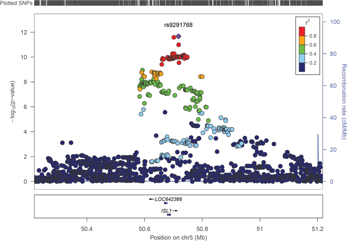
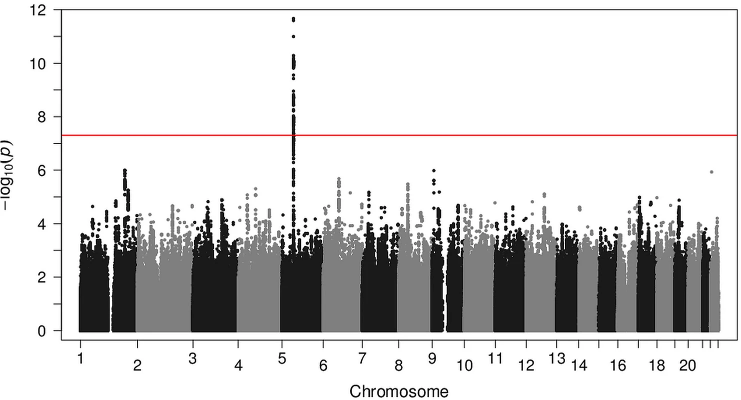
Next, we combined the effect estimates of GWAS1 and GWAS2 in a fixed effect meta-analysis. This meta-analysis also implicated the 220 kb region on chromosome 5q11.1. In the meta-analysis, multiple markers in this region showed evidence for association with CBE (Fig. 1). The most significant marker, rs9291768, had a P value of 2.13 x 10⁻¹². The possible relevance of rs9291768 in CBE was supported by the presence of 137 surrounding markers with P values of < 5 x 10⁻⁸. No other locus in the meta-analysis achieved this level of significance (Fig. 2). All markers with P values of < 10−5 are listed in S2 Table.
Fig. 1. Regional association plot for ISL1 across a 1.0 Mb window. 
Association with classic bladder exstrophy of individual SNPs in the meta-analysis GWAS is plotted as −log10(p) against chromosomal position. The y-axis on the right shows the recombination rate estimated from the 1000 Genomes (Mar 2012) EUR populations. All P values (y-axis on the left) are from the meta-analysis. The purple diamond indicates the most significant marker. Fig. 2. Genome-wide association scan in classic bladder exstrophy patients. 
Association of SNPs is plotted as −log10(p) against chromosomal position. The y-axis shows the negative log10 P values of the logistic regression for SNPs from the meta-analysis that passed quality control. Chromosomes are shown in alternating colors along the x-axis. The genome-wide significance level is indicated by a red line. The genotype-specific relative risks (RRs) for allele T of rs9291768 were: (i) RR_het = 2.00 for heterozygotes (95%-CI = 1.33–3.02); and (ii) RR_hom = 4.77 (95%-CI = 3.06–7.45) for homozygotes. This is compatible with neither a recessive (P = 3.9 x 10⁻⁵), nor a dominant mode of inheritance (P = 1.6 x 10⁻⁴). According to the genotype data from Ensembl release 74—December 2013©, the frequency of the CBE allele T at rs9291768 is highest in African populations (0.534), intermediate in European populations (0.425), and lowest in Asian populations (0.083).
Our previous study [20] failed to identify the possible relevance of both marker rs9291768 and the region comprising ISL1. In that report, rs9291768 obtained a P value of 1.1 x 10⁻³, which is not considered worthy of note in the context of a GWAS. In the present meta-analysis, the estimated relative risk for this SNP was 2.18 (see Table 1). The GWAS1 sample comprised 98 cases and 526 controls, and the power to achieve genome-wide significance (i.e. < 5 x 10⁻⁸) for a SNP with RR = 2.18 was only 31% under the assumption of a multiplicative model and a minor allele frequency of 0.377. For GWAS2, which comprised 110 cases and 1177 controls, the power was higher at 53%. However, the combination of GWAS1 and GWAS2 provides a power of 98%. This substantial increase in power is the central motivation for conducting meta-analyses. We therefore assume that the non-identification of rs9291768 in GWAS1 was attributable to the issues of power and random sample variation.
Tab. 1. Most strongly associated SNP in the bladder exstrophy susceptibility locus 5q11.1. 
Relative risks (RRs) are given with the risk allele set as baseline. Chr, chromosome; RAF, risk allele frequency. The marker rs9291768 is a non-coding variant, which is located 27.2 kb downstream of the ISL1 gene. The associated 220 kb haplotype block contains the gene ISL1. The only other transcript encoded in the regions flanking rs9291768 (500 kb on either side) is LOC642366, which also resides within the associated haplotype block. LOC642366 encodes an uncharacterized non-coding RNA, which has no ortholog in mouse, zebrafish, drosophila, C. elegans, or S. cerevisiae. The second most proximal gene to rs9291768 is Homo sapiens poly (ADP-ribose) polymerase family, member 8 (PARP8), which is located 575 kb proximal to rs9291768. The third and fourth genes are Homo sapiens pelota homolog (Drosophila) (PELO), and the integrin, alpha 1 subunit of integrin receptors (ITGA1), which are both located ∼1.4 Mb distal to rs9291768. According to Mouse Genome Informatics (http://www.informatics.jax.org/), neither PARP8 nor ITGA1 is expressed in the genital tubercle or the cloacal membrane during the CBE critical time frame in mouse embryos. Furthermore, mice with complete invalidation of PARP8 or ITGA1 display neither CBE features nor CBE-related phenotypes. Studies of the Drosophila pelota gene have implicated dPelo in spermatogenesis, mitotic division, and patterning. Homozygous Pelo-null embryos fail to develop beyond embryonic day 7.5, and exhibit no early CBE-related features, such as diastases of the symphysis. Whether rs9291768 per se, or a variant in linkage disequilibrium with it, confers the functional effect underlying the association remains unclear. The rs9291768 marker shows no association with any predicted regulatory sequence (according to ENCODE, TFSEARCH, or FAS-ESS), or splicing motif. Of the other 136 markers at this chromosome 5 locus, only one (rs2303751) is located in a coding region, and none affect a splice site. Marker rs2303751 is in linkage disequilibrium (r2 = 0.932) with the most significant marker rs9291768, and represents a synonymous A/G substitution in exon four of ISL1. Furthermore, none of the public eQTL (expression quantitative trait loci) databases contains evidence to suggest that rs9291768, or a SNP in perfect linkage disequilibrium with it, would affect gene expression levels (RegulomeDB, http://regulome.stanford.edu; eQTL browser, eqtl.uchicago.edu).
The present meta-analysis generated no evidence in support of the (non-genome-wide significant) association between CBE and an intergenic region on chromosome 17q21.31-q21.32 identified in GWAS1 [20]. This region is located between the genes WNT3 (wingless-type MMTV integration site family, member 3) and WNT9b (wingless-type MMTV integration site family, member 9b).
Since the CBE-associated region harbors the gene ISL1, we performed ISL1 re-sequencing in 207 CBE patients included in the present meta-analysis. As well as allowing mutation detection, this approach should provide genotype data for polymorphisms in the exons and exon-flanking regions of ISL1. Using the results of Sanger sequencing, we compared genotype information from four SNPs with the imputed data. We calculated the allelic accuracy, i.e. the aggregate difference between the actual number of alleles observed and the number of imputed alleles [21]. This yielded accuracy values of 96.9% (rs150104955); 97.3% (rs2288468); 97.3% (rs2303751); and 99.5% (rs3917084). Two of these SNPs (rs2288468, rs2303751) achieved genome-wide significance in the meta-analysis
Although sequencing identified no nonsense or probably pathogenic ISL1 variant, the following variants were all detected in a heterozygote state in single patients: intron 3, rs2303750; synonymous in exon 5, rs41268419 (p.Ser275 =); non-synonymous in exon 4, rs200209474 (p.Thr181Ser); unreported variants in intron 4, +21delG, -19delT, and -64A>G. Pathogenicity prediction using several publicly available algorithms (SNPs&Go, MutPred, SIFT) predicted that the p.Thr181Ser variant is neutral. Only PolyPhen-2 estimated it as possibly damaging. Furthermore, all of the observed intronic variants can be assumed to be benign. Hence, our patient sample size may have been too small to detect rare causal mutational events. We cannot exclude the possibility that some mutations were overlooked, i.e. mutations located in the promoter region, in as-yet-unknown regulatory sequences, or in non-coding regions that were not present within the covered sequence.
ISL1 encodes the insulin gene enhancer protein ISL1, a LIM zinc-binding/homeobox-domain transcription factor which was initially identified as a regulator of insulin expression [22]. Research in rodents suggests that Isl1 plays a fundamental role in the embryogenesis of multiple tissue types: Isl1 affects cell differentiation and survival, cell fate determination, the generation of cell diversity, and segmental patterning during mouse development [23]. Isl1 binds and regulates the promoters of the glucagon and somatostatin genes, and activates insulin gene transcription in pancreatic beta cells in synergy with NEUROD1 (neuronal differentiation 1) [24]. A previous study found an association between a heterozygous ISL1 premature termination mutation (p.Gln310*) and diabetes type II in a large Japanese kindred [25]. Furthermore, in a classic linkage analysis of 186 Swedish multiplex families with diabetes type I, linkage was observed with chromosomal region 5q11-q13, which harbors ISL1 [26]. This finding supports the hypothesis that ISL1 is implicated in pancreatic function and development, as reported in Isl1 knockout mice [27]. In the mouse, research at E (embryonic day) 8.5 to E9.5 has shown that ISL1 acts upstream of the sonic hedgehog (Shh) signaling pathway [28], which may be involved in other processes besides the coordination of heart and lung co-development [29]. Interestingly, a recent report by Matsumaru et al. [30] showed that SHH is also important for ventral body wall formation, and that ectopic SHH signaling induces omphalocele, a feature which is associated with CE, the severest form of the BEEC.
A previous study in mice also showed that a homozygous Isl1 null mutation (Isl1-/-) induced growth retardation at E9.5 and severe cardiac malformations at E10.5 [31]. Embryos exhibiting these severe cardiac malformations at E10.5 died at E11.5 due to the developmental arrest of spinal motor neurons [32]. Research has demonstrated a further role for Isl1 in mice at E11.5, i.e. in hindlimb-specific patterning and growth in combination with both SHH and the helix-loop-helix transcription factor HAND2 (heart and neural crest derivatives expressed 2) [33]. This interplay is also necessary for normal cardiac development in mice [23]. Recently, Jurberg et al. reported that specific activity of mouse Isl1 in the progenitors of the ventral lateral mesoderm promotes formation of the cloaca-associated mesoderm as the most posterior derivatives of lateral mesoderm progenitors [34]. This observation provides independent evidence that ISL1 is a promising candidate gene for human CBE.
In a recent mouse study, Kaku et al. induced conditional Isl1 deletion in the lateral mesoderm using a Hoxb6-Cre driver, and demonstrated that this caused kidney agenesis or hydroureter [35]. The authors observed transient Isl1 expression between E10.5—E14.5. At early stages, this was observed in the mesenchyme surrounding the ureteric stalk and cloaca. At later stages, expression occurred along the nephric duct, at the base of the ureteric stalk, and in the genital tubercle. This suggests that Isl1 may be implicated in kidney, ureter, and bladder development. These mice show a variable phenotype, which can include agenesis of the genital tubercle (R. Nishinakamura, personal communication). The variability of this defect is probably due to mosaicism, which arises as a result of the Hoxb6-Cre driver [33]. Kaku et al. also reported that conditional loss of Isl1 resulted in a concomitant reduction in the expression of bone morphogenetic protein 4 (Bmp4) [35]. Using mouse Isl1Cre;Bmp4flox/flox mutants, Suzuki et al. showed that BMP4 signaling in the caudal Isl1 expression domain was required for formation of the anterior peri-cloacal mesenchyme (aPCM) at E10.5 [36]. Rather than decreasing Isl1 function, loss of this signal caused defective pelvic and urogenital organ formation, including kidney and bladder agenesis, with abnormal development of the lower limbs and pelvis. Moreover, tissue lineage analyses suggested that Isl1-expressing cells are an essential cell population in terms of caudal body formation, including the pelvic/urogenital organs and hindlimb [36].
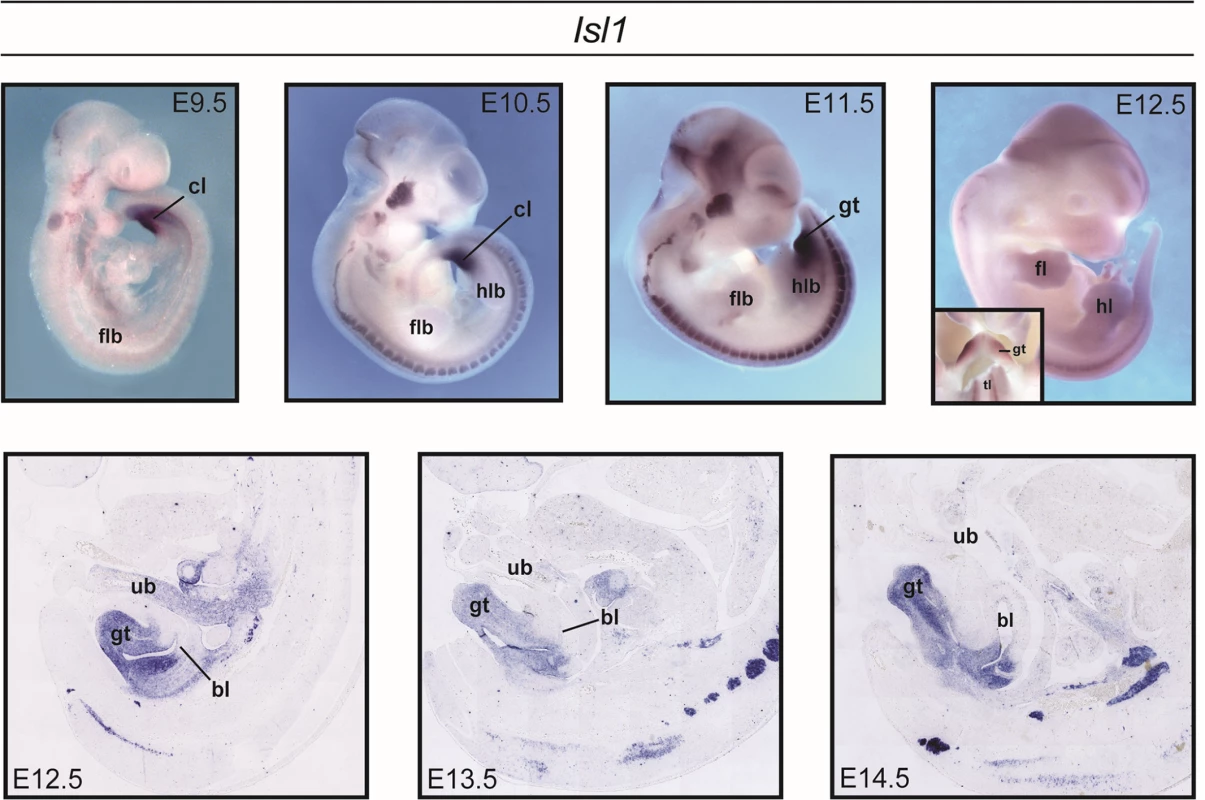
In the present mouse analyses, Isl1 was expressed during the critical timeframe for development of tissues involved in CBE, and strong Isl1 expression was detected in the developing genital region (Fig. 3). From E9.5, a broad Isl1 domain was detected in the cloacal region. This was maintained in the outgrowing genital tubercle (including the urorectal septum) until at least E14.5.
Fig. 3. Expression of Isl1 during mouse development. 
Whole-mount in situ hybridization (ISH) for Isl1 in wildtype mouse embryos between E9.5-E12.5 revealed strong expression in the developing genital region, including the cloaca, cloacal membrane, and emerging genital tubercle. ISH on mid-sagittal paraffin sections at later embryonic stages (E12.5-E14.5) revealed expression throughout the genital tubercle, within the peri-cloacal mesenchyme and urorectal septum. Isl1 was also detected in the craniofacial- and spinal ganglia. Three groups have recently elucidated the molecular basis of the Danforth’s short tail (Sd) mouse. They reported the insertion of a retrotransposon in the 5’ regulatory domain of the murine Ptf1a gene, which encodes pancreas specific transcription factor 1A [37–39]. As a consequence, and in contrast to their wildtype littermates, Sd mice showed ectopic Ptf1a expression in the notochord and hindgut at E8.5 to E9.5, which extended to the cloaca and mesonephros at E10.5 and to the pancreatic bud at E10.5 and E11.5 [38]. The resultant phenotype of this Sd mutation mirrors the phenotype observed in human caudal malformation syndromes, a phenotype that is also observed in Isl1 transgenic mice [40]. Moreover, the BEEC related human Currarino syndrome (MIM: #176450), which comprises hemisacrum, anorectal malformations, and a presacral mass, is caused by mutations of the transcription factor MNX1/HLXB9 (Motor neuron and pancreas homeobox protein 1). The genes Ptf1a, Isl1, and Mnx1 have been implicated in pancreas development, and MNX1 has been identified as a direct target of PTF1a [41].
Coordinated development of caudal body structures is necessary for the formation of the bladder, rectum, and the external genitalia [42,43]. These organs are derived from the transient embryonic cloaca and the PCM, an infra-umbilical mesenchyme [44], as well as the anterior PCM [36,42]. SHH-, ISL1-, and BMP4-expressing cells contribute to both the PCM and the anterior PCM [36,42,45]. Perturbation of this morphoregulatory network may thus lead to malformation of caudal structures, including the bladder, rectum, and external genitalia.
In summary, the present report describes a novel association between ISL1 and human CBE. While previous conventional linkage - and candidate gene studies in humans have suggested the involvement of ISL1 in diabetes type I and II and in congenital heart defects, to our knowledge, the present study is the first to implicate ISL1 in the formation of human urogenital malformations. The observed variation in CBE birth prevalence across populations is consistent with the cross-population frequencies of the rs9291768 T-allele, thus supporting our finding.
The importance of Pft1a and Isl1 in the formation of murine genital development and caudal regression phenotypes, the involvement of MNX1 in the BEEC related human Currarino syndrome, and the role of all three genes in pancreatic development suggest that these genes are involved in a common pathway. However, the present data do not exclude the possibility that the association between CBE and the region surrounding ISL1 is attributable to long-range functional interactions with other regions in the human genome. Future studies are warranted to identify the mechanisms through which genetic variation at ISL1 contributes to CBE development.
Methods
Subjects GWAS2
The initial GWAS2 sample comprised 123 isolated CBE patients and 1,320 controls of European descent. Prior to inclusion, written informed consent was obtained from all subjects, or from their proxies in the case of legal minors. For patients and controls, demographic information was collected using a structured questionnaire. This study was approved by the institutional ethics committee of each participating center, and was conducted in accordance with the principles of the Declaration of Helsinki. All CBE patients were recruited in person by experienced physicians trained in the diagnosis of the BEEC. Details of the recruitment process for patients and controls are provided in Reutter et al. [20].
Genotyping
For the 123 isolated CBE patients in GWAS2, genotyping was performed using the Illumina BeadChip HumanOmniExpress (San Diego, California, USA), and DNA was extracted from blood or saliva using standard procedures. Case-control comparisons were made using the genotypes of 1,320 population-based controls, which had been processed using the same array [46]. Genome-wide genotyping of 730,525 markers was conducted using the Infinium HD Ultra Assay from Illumina (Illumina, San Diego, California, USA).
Pre-imputation quality control of GWAS2
Markers were excluded from the analysis if: (i) the minor allele frequency was <1% or the call rate was <95% in either cases or controls; or (ii) the test for Hardy-Weinberg equilibrium resulted in P<10−4 in the control sample or P<10−6 in the case sample. A total of 616,799 autosomal markers fulfilled these quality criteria. Individuals were excluded if their call rate was <99%, or if they were outliers in a multidimensional scaling (MDS) analysis. Relatedness of individuals within GWAS2, and between GWAS1 and GWAS2, was evaluated using both the KING program [47], and an identity-by-state-based in-house program. The post quality control data set of GWAS2 comprised 110 CBE patients and 1,177 controls.
Imputation
GWAS1 and GWAS2 were imputed separately to the 1000 Genomes Project and HapMap 3 reference panels using IMPUTE2 [48].
Post-imputation quality control
For each of the three data sets, variants were excluded if: (i) the imputation info score was <0.4; (ii) the dosage of the minor allele was <1% in either cases or controls; (iii) the test for Hardy-Weinberg equilibrium (calculated on the basis of the 80% best-guess genotypes) resulted in P<10−4 in the control sample; or (iv) the 80% best-guess genotypes were only available for <80% of cases or controls. In total, 7,261,187 SNPs were analyzed in at least one data set.
Statistical analysis
Single-marker analysis was performed using logistic regression. The allele dosage and the first five components obtained from MDS were used as independent variables for the variants in the three data sets. The effect estimates for the data sets were then combined in an inverse variance-weighted fixed-effects meta-analysis. The genomic inflation factor in this meta-analysis was 1.0196.
Power
Power was calculated to enable detection of genome-wide significance (P <5 x 10⁻⁸) in the combined analysis of the GWAS1 and GWAS2 samples. Under the assumption of a multiplicative model, this was 80% for an allele frequency of 0.35 (0.20) and a RR of 1.94 (2.05). This is within the range of RRs observed for other multifactorial, nonsyndromic human malformations. For example, the power of the present study to detect a locus with an effect-strength similar to that of the most strongly associated locus in nonsyndromic cleft lip with or without cleft palate was 99.2% [49].
ISL-1 resequencing
Sequence analysis of the complete ISL-1 coding regions and their splice consensus motifs was performed in 207 of our 208 CBE patients using standard techniques. Primers are listed in S3 Table. For the remaining patient, no additional DNA sample was available. During this analysis, we also obtained information for several SNPs deposited in dbSNP Build142 (rs3917084, rs150104955, rs2288468, rs2303750, rs2303751, rs200209474, and rs41268419).
In situ hybridization of mouse embryo sections
The expression of Isl1 was analyzed using in situ hybridization, standard procedures, and a ∼450bp antisense probe spanning exons 2 and 3 from XM_006517533.1. Details of the in situ hybridization methods are provided elsewhere [50].
Supporting Information
Zdroje
1. Husmann DA, Vandersteen DR (1999) Anatomy of the cloacal exstrophy. In: Gearhart JP, Mathews R (eds.): The exstrophy-epispadias complex, New York, Kluwer Academic/Plenum Publishers, pp. 199–206.
2. Hurst JA (2012) Anterior abdominal wall defects,” In: Firth HV, Hall JG (eds.): Oxford desk reference. Clinical genetics, New York, Oxford University Press, p.566.
3. Stein R, Fisch M, Black P, Hohenfellner R (1999) Strategies for reconstruction after unsuccessful or unsatisfactory primary treatment of patients with bladder exstrophy or incontinent epispadias. J Urol 161 : 1934–1941. 10332476
4. Ebert AK, Reutter H, Ludwig M, Rösch WH (2009) The exstrophy-epispadias complex. Orphanet J Rare Dis 4 : 1–17. doi: 10.1186/1750-1172-4-1 19133130
5. Reutter H, Qi L, Gearhart JP, Boemers T, Ebert AK, et al. (2007) Concordance analyses of twins with bladder exstrophy-epispadias complex suggest genetic etiology. Am J Med Genet A 143A: 2751–2756. 17937426
6. Gearhart JP, Jeffs RD (1998) Exstrophy-epispadias complex and bladder anomalies. In Campbell’s Urology. 7th edition, Walsh P.C., Retik A.B., Vaughan E.D. and Wein A,J, ed. (Philadelphia, US: WB Saunders), pp. 1939–1990.
7. Wiesel A, Queisser-Luft A, Clementi M, Bianca S, Stoll C (2005) Prenatal detection of congenital renal malformations by fetal ultrasonographic examination: an analysis of 709,030 births in 12 European countries. Eur J Med Genet 48 : 131–144. 16053904
8. International Clearinghouse for Birth Defects Monitoring Systems (1987) Epidemiology of bladder exstrophy and epispadias: a communication from the International Clearinghouse for Birth Defects Monitoring Systems. Teratology 36 : 221–227. 3424208
9. Boyadjiev SA, Dodson JL, Radford CL, Ashrafi GH, Beaty TH, et al. (2004) Clinical and molecular characterization of the bladder exstrophy-epispadias complex: analysis of 232 families. BJU Int 94 : 1337–1343. 15610117
10. Reutter H, Boyadjiev SA, Gambhir L, Ebert AK, Rösch W, et al. (2011) Phenotype severity in the bladder exstrophy-epispadias complex: analysis of genetic and nongenetic contributing factors in 441 families from North America and Europe. J Pediatr 159 : 825–831. doi: 10.1016/j.jpeds.2011.04.042 21679965
11. Shapiro E, Lepor H, Jeffs RD (1984) The inheritance of the exstrophy-epispadias complex. J Urol 132 : 308–310. 6737583
12. Messelink EJ, Aronson DC, Knuist M, Heij HA, Vos A (1994) Four cases of bladder exstrophy in two families. J Med Genet 31 : 490–492. 8071977
13. Ludwig M, Ching B, Reutter H, Boyadjiev SA (2009) The bladder exstrophy-epispadias complex. Birth Defects Res Part A Clin Mol Teratol 85 : 509–522. doi: 10.1002/bdra.20557 19161161
14. Reutter H, Shapiro E, Gruen JR (2003) Seven new cases of familial isolated bladder exstrophy and epispadias complex (BEEC) and review of the literature. Am J Med Genet A 120A:215–221. 12833402
15. Draaken M, Reutter H, Schramm C, Bartels E, Boemers TM, et al. (2010) Microduplications at 22q11.21 are associated with non-syndromic classic bladder exstrophy. Eur J Med Genet 53 : 55–60. doi: 10.1016/j.ejmg.2009.12.005 20060941
16. Lundin J, Söderhäll C, Lundén L, Hammarsjö A, White I, et al. (2010) 22q11.2 microduplication in two patients with bladder exstrophy and hearing impairment. Eur J Med Genet 53 : 61–65. doi: 10.1016/j.ejmg.2009.11.004 20045748
17. Draaken M, Baudisch F, Timmermann B, Kuhl H, Kerick M, et al. (2014) Classic bladder exstrophy: Frequent 22q11.21 duplications and definition of a 414 kb phenocritical region. Birth Defects Res Part A Clin Mol Teratol 100 : 512–517. doi: 10.1002/bdra.23249 24764164
18. Wilkins S, Zhang KW, Mahfuz I, Quantin R, D'Cruz N, et al. (2012) Insertion/deletion polymorphisms in the ΔNp63 promoter are a risk factor for bladder exstrophy epispadias complex. PLoS Genet 8: e1003070. doi: 10.1371/journal.pgen.1003070 23284286
19. Qi L, Wang M, Yagnick G, Gearhart JP, Ebert AK, et al. (2013) Candidate gene association study implicates p63 in the etiology of nonsyndromic bladder-exstrophy-epispadias complex. Birth Defects Res Part A Clin Mol Teratol 97 : 759–763. doi: 10.1002/bdra.23161 23913486
20. Reutter H, Draaken M, Pennimpede T, Wittler L, Brockschmidt FF, et al. (2014) Genome-wide association study and mouse expression data identify a highly conserved 32 kb intergenic region between WNT3 and WNT9b as possible susceptibility locus for isolated classic exstrophy of the bladder. Hum Mol Genet (in press), doi: 10.1093/hmg/ddu259
21. Raychaudhuri S, Sandor C, Stahl EA, Freudenberg J, Lee HS, et al. (2012) Five amino acids in three HLA proteins explain most of the association between MHC and seropositive rheumatoid arthritis. Nat Genet 44 : 291–296. doi: 10.1038/ng.1076 22286218
22. Karlsson O, Thor S, Norberg T, Ohlsson H, Edlund T (1990) Insulin gene enhancer binding protein Isl-1 is a member of a novel class of proteins containing both a homeo - and a Cys-His domain. Nature 344 : 879–882. 1691825
23. Zhuang S, Zhang Q, Zhuang T, Evans SM, Liang X, Sun Y (2013) Expression of Isl1 during mouse development. Gene Expr Patterns 13 : 407–412. doi: 10.1016/j.gep.2013.07.001 23906961
24. Zhang H, Wang WP, Guo T, Yang JC, Chen P, et al. (2009) The LIM-homeodomain protein ISL1 activates insulin gene promoter directly through synergy with BETA2. J Mol Biol 392 : 566–577. doi: 10.1016/j.jmb.2009.07.036 19619559
25. Shimomura H, Sanke T, Hanabusa T, Tsunoda K, Furuta H, Nanjo K (2000) Nonsense mutation of islet-1 gene (Q310X) found in a type 2 diabetic patient with a strong family history. Diabetes 49 : 1597–1600. 10969846
26. Holm P, Rydlander B, Luthman H, Kockum I, European Consortium for IDDM Genome Studies (2004) Interaction and association analysis of a type 1 diabetes susceptibility locus on chromosome 5q11-q13 and the 7q32 chromosomal region in Scandinavian families. Diabetes 53 : 1584–1591. 15161765
27. Ahlgren U, Pfaff SL, Jessell TM, Edlund T, Edlund H (1997) Independent requirement for ISL1 in formation of pancreatic mesenchyme and islet cells. Nature 385 : 257–260. 9000074
28. Lin L, Bu L, Cai CL, Zhang X, Evans S (2013) Isl1 is upstream of sonic hedgehog in a pathway required for cardiac morphogenesis. Dev Biol 295 : 756–763.
29. Peng T, Tian Y, Boogerd CJ, Lu MM, Kadzik RS, et al. (2013) Coordination of heart and lung co-development by a multipotent cardiopulmonary progenitor. Nature 500 : 589–593. doi: 10.1038/nature12358 23873040
30. Matsumaru D, Haraguchi R, Miyagawa S, Motoyama J, Nakagata N, Meijlink F, Yamada G (2011) Genetic analysis of hedgehog signaling in ventral body wall development and the onset of omphalocele formation. PLoS ONE 6: e16260. doi: 10.1371/journal.pone.0016260 21283718
31. Cai CL, Liang X, Shi Y, Chu PH, Pfaff SL, Chen J, Evans S (2003) Isl1 identifies a cardiac progenitor population that proliferates prior to differentiation and contributes a majority of cells to the heart. Dev Cell 5 : 877–889. 14667410
32. Pfaff SL, Mendelsohn M, Stewart CL, Edlund T, Jessell TM (1996) Requirement for LIM homeobox gene Isl1 in motor neuron generation reveals a motor neuron-dependent step in interneuron differentation. Cell 84 : 309–320. 8565076
33. Itou J, Kawakami H, Quach T, Osterwalder M, Evans SM, Zeller R, Kawakami Y (2012) Islet1 regulates establishment of the posterior hindlimb field upstream of the Hand2-Shh morphoregulatory gene network in mouse embryos. Development 139 : 1620–1629. doi: 10.1242/dev.073056 22438573
34. Jurberg AD, Aires R, Varela-Lasheras I, Nóvoa A, Mallo M (2013) Switching axial progenitors from producing trunk to tail tissues in vertebrate embryos. Dev Cell 25 : 451–462. doi: 10.1016/j.devcel.2013.05.009 23763947
35. Kaku Y, Ohmori T, Kudo K, Fujimura S, Suzuki S, et al. (2013) Islet1 deletion causes kidney agenesis and hydroureter resembling CAKUT. J Am Soc Nephrol 24 : 1242–1249. doi: 10.1681/ASN.2012050528 23641053
36. Suzuki K, Adachi Y, Numata T, Nakada S, Yanagita M, et al. (2012) Reduced BMP signaling results in hindlimb fusion with lethal pelvic/urogenital organ aplasia: a new mouse model of sirenomelia. PLoS ONE 7: e43453. doi: 10.1371/journal.pone.0043453 23028455
37. Lugani F, Arora R, Papeta N, Patel A, Zheng Z, et al. (2013) A retrotransposon insertion in the 5’ regulatory domain of Ptf1a results in ectopic gene expression and multiple congenital defects in Danforth’s short tail mouse. PLoS Genet 9: e1003206. doi: 10.1371/journal.pgen.1003206 23437001
38. Semba K, Araki K, Matsumoto K, Suda H, Ando T, et al. (2013) Ectopic expression of Ptf1a induces spinal defects, urogenital defects, and anorectal malformations in Danforth’s short tail mice. PLoS Genet 9: e1003204. doi: 10.1371/journal.pgen.1003204 23436999
39. Vlangos CN, Siuniak AN, Robinson D, Chinnaiyan AM, Lyons RH Jr, Cavalcoli JD, Keegan CE (2013) Next-generation sequencing identifies the Danforth’s short tail mouse mutation as a retrotransposon insertion affecting Ptf1a expression. PLoS Genet 9: e1003205. doi: 10.1371/journal.pgen.1003205 23437000
40. Muller YL, Yueh YG, Yaworsky PJ, Salbaum JM, Kappen C (2003) Caudal dysgenesis in Islet-1 transgenic mice. FASEB J 17 : 1349–1351. 12738808
41. Thompson N, Gésina E, Scheinert P, Bucher P, Grapin-Botton A (2012) RNA profiling and chromatin immunoprecipitation-sequencing reveal that PTF1a stabilizes pancreas progenitor identity via the control of MNX1/HLXB9 and a network of other transcription factors. Mol Cell Biol 32 : 1189–1199. doi: 10.1128/MCB.06318-11 22232429
42. Haraguchi R, Motoyama J, Sasaki H, Satoh Y, Miyagawa S, et al. (2007) Molecular analysis of coordinated bladder and urogenital organ formation by Hedgehog signaling. Development 134 : 525–533. 17202190
43. Haraguchi R, Matsumaru D, Nakagata N, Miyagawa S, Suzuki K, Kitazawa S, Yamada G (2012) The hedgehog signal induced modulation of bone morphogenetic protein signaling: an essential signaling relay for urinary tract morphogenesis. PLoS ONE 7: e42245. doi: 10.1371/journal.pone.0042245 22860096
44. Mildenberger H, Kluth D, Dziuba M (1988) Embryology of bladder exstrophy. J Pediatr Surg 23 : 166–170. 3343652
45. Haraguchi R, Mo R, Hui C, Motoyama J, Makino S, et al. (2001) Unique functions of Sonic hedgehog signaling during external genitalia development. Development 128 : 4241–4250. 11684660
46. Schmermund A, Möhlenkamp S, Stang A, Grönemeyer D, Seibel R, et al. (2002) Assessment of clinically silent atherosclerotic disease and established and novel risk factors for predicting myocardial infarction and cardiac death in healthy middle-aged subjects: rationale and design of the Heinz Nixdorf RECALL Study. Risk Factors, Evaluation of Coronary Calcium and Lifestyle. Am Heart J 144 : 212–218. 12177636
47. Manichaikul A, Mychaleckyj JC, Rich SS, Daly K, Sale M, Chen WM (2010) Robust relationship inference in genome-wide association studies. Bioinformatics 26 : 2867–2873. doi: 10.1093/bioinformatics/btq559 20926424
48. Howie BN, Donnelly P, Marchini J (2009) A flexible and accurate genotype imputation method for the next generation of genome-wide association studies. PLoS Genet 5: e1000529. doi: 10.1371/journal.pgen.1000529 19543373
49. Birnbaum S, Ludwig KU, Reutter H, Herms S, Steffens M, et al. (2009) Key susceptibility locus for nonsyndromic cleft lip with or without cleft palate on chromosome 8q24. Nat Genet 41 : 473–477. doi: 10.1038/ng.333 19270707
50. Pennimpede T, Proske J, König A, Vidigal JA, Morkel M, et al. (2012) In vivo knockdown of Brachyury results in skeletal defects and urorectal malformations resembling caudal regression syndrome. Dev Biol 372 : 55–67. doi: 10.1016/j.ydbio.2012.09.003 22995555
Štítky
Genetika Reprodukčná medicína
Článek NLRC5 Exclusively Transactivates MHC Class I and Related Genes through a Distinctive SXY ModuleČlánek Inhibition of Telomere Recombination by Inactivation of KEOPS Subunit Cgi121 Promotes Cell LongevityČlánek HOMER2, a Stereociliary Scaffolding Protein, Is Essential for Normal Hearing in Humans and MiceČlánek LRGUK-1 Is Required for Basal Body and Manchette Function during Spermatogenesis and Male FertilityČlánek The GATA Factor Regulates . Developmental Timing by Promoting Expression of the Family MicroRNAsČlánek Systems Biology of Tissue-Specific Response to Reveals Differentiated Apoptosis in the Tick VectorČlánek Phenotype Specific Analyses Reveal Distinct Regulatory Mechanism for Chronically Activated p53Článek The Nuclear Receptor DAF-12 Regulates Nutrient Metabolism and Reproductive Growth in NematodesČlánek The ATM Signaling Cascade Promotes Recombination-Dependent Pachytene Arrest in Mouse SpermatocytesČlánek The Small Protein MntS and Exporter MntP Optimize the Intracellular Concentration of Manganese
Článok vyšiel v časopisePLOS Genetics
Najčítanejšie tento týždeň
2015 Číslo 3- Gynekologové a odborníci na reprodukční medicínu se sejdou na prvním virtuálním summitu
- Je „freeze-all“ pro všechny? Odborníci na fertilitu diskutovali na virtuálním summitu
-
Všetky články tohto čísla
- NLRC5 Exclusively Transactivates MHC Class I and Related Genes through a Distinctive SXY Module
- Licensing of Primordial Germ Cells for Gametogenesis Depends on Genital Ridge Signaling
- A Genomic Duplication is Associated with Ectopic Eomesodermin Expression in the Embryonic Chicken Comb and Two Duplex-comb Phenotypes
- Genome-wide Association Study and Meta-Analysis Identify as Genome-wide Significant Susceptibility Gene for Bladder Exstrophy
- Mutations of Human , Encoding the Mitochondrial Asparaginyl-tRNA Synthetase, Cause Nonsyndromic Deafness and Leigh Syndrome
- Exome Sequencing in an Admixed Isolated Population Indicates Variants Confer a Risk for Specific Language Impairment
- Genome-Wide Association Studies in Dogs and Humans Identify as a Risk Variant for Cleft Lip and Palate
- Rapid Evolution of Recombinant for Xylose Fermentation through Formation of Extra-chromosomal Circular DNA
- The Ribosome Biogenesis Factor Nol11 Is Required for Optimal rDNA Transcription and Craniofacial Development in
- Methyl Farnesoate Plays a Dual Role in Regulating Metamorphosis
- Maternal Co-ordinate Gene Regulation and Axis Polarity in the Scuttle Fly
- Maternal Filaggrin Mutations Increase the Risk of Atopic Dermatitis in Children: An Effect Independent of Mutation Inheritance
- Inhibition of Telomere Recombination by Inactivation of KEOPS Subunit Cgi121 Promotes Cell Longevity
- Clonality and Evolutionary History of Rhabdomyosarcoma
- HOMER2, a Stereociliary Scaffolding Protein, Is Essential for Normal Hearing in Humans and Mice
- Methylation-Sensitive Expression of a DNA Demethylase Gene Serves As an Epigenetic Rheostat
- BREVIPEDICELLUS Interacts with the SWI2/SNF2 Chromatin Remodeling ATPase BRAHMA to Regulate and Expression in Control of Inflorescence Architecture
- Seizures Are Regulated by Ubiquitin-specific Peptidase 9 X-linked (USP9X), a De-Ubiquitinase
- The Fun30 Chromatin Remodeler Fft3 Controls Nuclear Organization and Chromatin Structure of Insulators and Subtelomeres in Fission Yeast
- A Cascade of Iron-Containing Proteins Governs the Genetic Iron Starvation Response to Promote Iron Uptake and Inhibit Iron Storage in Fission Yeast
- Mutation in MRPS34 Compromises Protein Synthesis and Causes Mitochondrial Dysfunction
- LRGUK-1 Is Required for Basal Body and Manchette Function during Spermatogenesis and Male Fertility
- Cis-Regulatory Mechanisms for Robust Olfactory Sensory Neuron Class-restricted Odorant Receptor Gene Expression in
- Effects on Murine Behavior and Lifespan of Selectively Decreasing Expression of Mutant Huntingtin Allele by Supt4h Knockdown
- HDAC4-Myogenin Axis As an Important Marker of HD-Related Skeletal Muscle Atrophy
- A Conserved Domain in the Scc3 Subunit of Cohesin Mediates the Interaction with Both Mcd1 and the Cohesin Loader Complex
- Selective and Genetic Constraints on Pneumococcal Serotype Switching
- Bacterial Infection Drives the Expression Dynamics of microRNAs and Their isomiRs
- The GATA Factor Regulates . Developmental Timing by Promoting Expression of the Family MicroRNAs
- Accumulation of Glucosylceramide in the Absence of the Beta-Glucosidase GBA2 Alters Cytoskeletal Dynamics
- Reproductive Isolation of Hybrid Populations Driven by Genetic Incompatibilities
- The Contribution of Alu Elements to Mutagenic DNA Double-Strand Break Repair
- Systems Biology of Tissue-Specific Response to Reveals Differentiated Apoptosis in the Tick Vector
- Tfap2a Promotes Specification and Maturation of Neurons in the Inner Ear through Modulation of Bmp, Fgf and Notch Signaling
- The Lysine Acetyltransferase Activator Brpf1 Governs Dentate Gyrus Development through Neural Stem Cells and Progenitors
- PHABULOSA Controls the Quiescent Center-Independent Root Meristem Activities in
- DNA Polymerase ζ-Dependent Lesion Bypass in Is Accompanied by Error-Prone Copying of Long Stretches of Adjacent DNA
- Examining the Evolution of the Regulatory Circuit Controlling Secondary Metabolism and Development in the Fungal Genus
- Zinc Finger Independent Genome-Wide Binding of Sp2 Potentiates Recruitment of Histone-Fold Protein Nf-y Distinguishing It from Sp1 and Sp3
- GAGA Factor Maintains Nucleosome-Free Regions and Has a Role in RNA Polymerase II Recruitment to Promoters
- Neurospora Importin α Is Required for Normal Heterochromatic Formation and DNA Methylation
- Ccr4-Not Regulates RNA Polymerase I Transcription and Couples Nutrient Signaling to the Control of Ribosomal RNA Biogenesis
- Phenotype Specific Analyses Reveal Distinct Regulatory Mechanism for Chronically Activated p53
- A Systems-Level Interrogation Identifies Regulators of Blood Cell Number and Survival
- Morphological Mutations: Lessons from the Cockscomb
- Genetic Interaction Mapping Reveals a Role for the SWI/SNF Nucleosome Remodeler in Spliceosome Activation in Fission Yeast
- The Role of China in the Global Spread of the Current Cholera Pandemic
- The Nuclear Receptor DAF-12 Regulates Nutrient Metabolism and Reproductive Growth in Nematodes
- A Zinc Finger Motif-Containing Protein Is Essential for Chloroplast RNA Editing
- Resistance to Gray Leaf Spot of Maize: Genetic Architecture and Mechanisms Elucidated through Nested Association Mapping and Near-Isogenic Line Analysis
- Small Regulatory RNA-Induced Growth Rate Heterogeneity of
- Mitochondrial Dysfunction Reveals the Role of mRNA Poly(A) Tail Regulation in Oculopharyngeal Muscular Dystrophy Pathogenesis
- Complex Genomic Rearrangements at the Locus Include Triplication and Quadruplication
- Male-Biased Aganglionic Megacolon in the TashT Mouse Line Due to Perturbation of Silencer Elements in a Large Gene Desert of Chromosome 10
- Sex Ratio Meiotic Drive as a Plausible Evolutionary Mechanism for Hybrid Male Sterility
- Tertiary siRNAs Mediate Paramutation in .
- RECG Maintains Plastid and Mitochondrial Genome Stability by Suppressing Extensive Recombination between Short Dispersed Repeats
- Escape from X Inactivation Varies in Mouse Tissues
- Opposite Phenotypes of Muscle Strength and Locomotor Function in Mouse Models of Partial Trisomy and Monosomy 21 for the Proximal Region
- Glycosyl Phosphatidylinositol Anchor Biosynthesis Is Essential for Maintaining Epithelial Integrity during Embryogenesis
- Hyperdiverse Gene Cluster in Snail Host Conveys Resistance to Human Schistosome Parasites
- The Class Homeodomain Factors and Cooperate in . Embryonic Progenitor Cells to Regulate Robust Development
- Recombination between Homologous Chromosomes Induced by Unrepaired UV-Generated DNA Damage Requires Mus81p and Is Suppressed by Mms2p
- Synergistic Interactions between Orthologues of Genes Spanned by Human CNVs Support Multiple-Hit Models of Autism
- Gene Networks Underlying Convergent and Pleiotropic Phenotypes in a Large and Systematically-Phenotyped Cohort with Heterogeneous Developmental Disorders
- The ATM Signaling Cascade Promotes Recombination-Dependent Pachytene Arrest in Mouse Spermatocytes
- Combinatorial Control of Light Induced Chromatin Remodeling and Gene Activation in
- Linking Aβ42-Induced Hyperexcitability to Neurodegeneration, Learning and Motor Deficits, and a Shorter Lifespan in an Alzheimer’s Model
- The Complex Contributions of Genetics and Nutrition to Immunity in
- NatB Domain-Containing CRA-1 Antagonizes Hydrolase ACER-1 Linking Acetyl-CoA Metabolism to the Initiation of Recombination during . Meiosis
- Transcriptomic Profiling of Reveals Reprogramming of the Crp Regulon by Temperature and Uncovers Crp as a Master Regulator of Small RNAs
- Osteopetrorickets due to Snx10 Deficiency in Mice Results from Both Failed Osteoclast Activity and Loss of Gastric Acid-Dependent Calcium Absorption
- A Genomic Portrait of Haplotype Diversity and Signatures of Selection in Indigenous Southern African Populations
- Sequence Features and Transcriptional Stalling within Centromere DNA Promote Establishment of CENP-A Chromatin
- Inhibits Neuromuscular Junction Growth by Downregulating the BMP Receptor Thickveins
- Replicative DNA Polymerase δ but Not ε Proofreads Errors in and in
- Unsaturation of Very-Long-Chain Ceramides Protects Plant from Hypoxia-Induced Damages by Modulating Ethylene Signaling in
- The Small Protein MntS and Exporter MntP Optimize the Intracellular Concentration of Manganese
- A Meta-analysis of Gene Expression Signatures of Blood Pressure and Hypertension
- Pervasive Variation of Transcription Factor Orthologs Contributes to Regulatory Network Evolution
- Network Analyses Reveal Novel Aspects of ALS Pathogenesis
- A Role for the Budding Yeast Separase, Esp1, in Ty1 Element Retrotransposition
- Nab3 Facilitates the Function of the TRAMP Complex in RNA Processing via Recruitment of Rrp6 Independent of Nrd1
- A RecA Protein Surface Required for Activation of DNA Polymerase V
- PLOS Genetics
- Archív čísel
- Aktuálne číslo
- Informácie o časopise
Najčítanejšie v tomto čísle- Clonality and Evolutionary History of Rhabdomyosarcoma
- Morphological Mutations: Lessons from the Cockscomb
- Maternal Filaggrin Mutations Increase the Risk of Atopic Dermatitis in Children: An Effect Independent of Mutation Inheritance
- Transcriptomic Profiling of Reveals Reprogramming of the Crp Regulon by Temperature and Uncovers Crp as a Master Regulator of Small RNAs
Prihlásenie#ADS_BOTTOM_SCRIPTS#Zabudnuté hesloZadajte e-mailovú adresu, s ktorou ste vytvárali účet. Budú Vám na ňu zasielané informácie k nastaveniu nového hesla.
- Časopisy


