-
Články
- Časopisy
- Kurzy
- Témy
- Kongresy
- Videa
- Podcasty
It's All in Your Mind: Determining Germ Cell Fate by Neuronal IRE-1 in
Cells in the C.
elegans germline undergo programmed cell death as part of the normal developmental program and in response to various stresses. Here, we discovered that more germ cells undergo programmed cell death under stress conditions associated with the accumulation of misfolded proteins in the endoplasmic reticulum, a cellular organelle responsible for protein folding and trafficking. Surprisingly, we found that germ cell death is a consequence of stress in neurons rather than in the germ cells themselves. This implies that germ cell death under ER stress conditions is regulated at the organismal level and implicates signaling between tissues.
Published in the journal: It's All in Your Mind: Determining Germ Cell Fate by Neuronal IRE-1 in. PLoS Genet 10(10): e32767. doi:10.1371/journal.pgen.1004747
Category: Research Article
doi: https://doi.org/10.1371/journal.pgen.1004747Summary
Cells in the C.
elegans germline undergo programmed cell death as part of the normal developmental program and in response to various stresses. Here, we discovered that more germ cells undergo programmed cell death under stress conditions associated with the accumulation of misfolded proteins in the endoplasmic reticulum, a cellular organelle responsible for protein folding and trafficking. Surprisingly, we found that germ cell death is a consequence of stress in neurons rather than in the germ cells themselves. This implies that germ cell death under ER stress conditions is regulated at the organismal level and implicates signaling between tissues.Introduction
Apoptosis, also known as programed cell death (PCD), is a highly conserved fundamental cellular process that provides a self-elimination mechanism for the removal of unwanted cells. PCD is critical for organ development, tissue remodeling, cellular homeostasis and elimination of abnormal and damaged cells [1], [2]. The apoptotic machinery that actually executes cell death is intrinsic to all cells and can be activated in response to extracellular or intracellular cues. These are thought to be mediated by cell death receptors or by cytotoxic stress respectively [3].
In C. elegans, 131 somatic cells invariably undergo apoptosis during hermaphrodite development [4], [5]. In contrast, in the adult C. elegans, only germ cells undergo apoptotic cell death. These cell deaths can be either physiological or stress-induced [6], [7]. So far, stress-induced germ cell apoptosis has been associated with DNA damage, pathogens, oxidative stress, osmotic stress, heat shock and starvation [7]–[9]. These apoptotic events are restricted to germ cells at the pachytene stage which are located in the loop region of the gonad [6], where oogenesis transition normally occurs [10]. The physiological germ cell apoptosis pathway acts during oogenesis and is thought to act either as a part of a quality control process, preferentially removing unfit germ cells from the gonad, or as a resource re-allocation factor important for maintaining oocyte quality or simply as a gonad homeostatic pathway removing excess germ cells [6], [11], [12]. Both somatic and germ cell apoptosis rely on the highly conserved core apoptotic machinery comprised of the Caspase-3 homolog ced-3, the Apaf-1 homolog ced-4 and the anti-apoptotic Bcl-2 homolog ced-9 [6], [13]–[16].
All germ cell apoptosis, physiological and stress-induced, relies on the core apoptotic machinery [7]–[9]. However, different upstream genes activate the core apoptotic machinery in the germline in response to different stresses. For example, DNA damage-induced germ cell apoptosis involves the proteins EGL-1, CED-13, and the DNA damage response protein p53 homolog CEP-1 [9], [17]–[19]. In contrast, oxidative, osmotic, heat shock and starvation stresses induce germ cell apoptosis through a CEP-1 and EGL-1 independent pathway and rely on the MEK-1 and SEK-1 MAPKs instead [7].
The endoplasmic reticulum (ER) fulfills many essential cellular functions, including a role in the secretory pathway, in lipid metabolism and in calcium sequestration. Accordingly, ER homeostasis is essential for proper cellular function [20]. A specialized, conserved cellular stress response, called the unfolded protein response (UPR), is in charge of detecting ER stress and adjusting the capacity of the ER to restore ER homeostasis. In C. elegans, as in humans, three proteins located at the ER membrane sense ER stress and activate the UPR: the ribonuclease inositol-requiring protein-1 (IRE-1), the PERK kinase homolog PEK-1 and the activating transcription factor-6 (ATF-6) [21]. Of the three, IRE-1 is the major and most highly conserved ER stress sensor. In response to ER stress, IRE-1 activates the ER stress-related transcription factor XBP-1, which induces the transcription of genes that help restore ER homeostasis [22]–[24]. Accordingly, ire-1 and xbp-1 deficiencies perturb ER homeostasis [25], [26].
Although IRE-1 typically protects cells, upon excessive and prolonged ER stress, IRE-1 can also trigger cell death, usually in the form of apoptosis [27], [28]. For example, IRE-1 can lead to activation of the cell death machinery via JNK and caspase activation [29], [30] or by mediating decay of critical ER-localized mRNAs through the RIDD pathway, tipping the balance in favor of apoptosis [31]. These functions of IRE-1 are independent of XBP-1 [29]–[33].
Highly proliferating cells with a high protein and lipid biosynthetic load are thought to rely on ER function to a greater extent than other cells. This together with the general sensitivity of the germline to cellular stresses prompted us to investigate the effects of ER stress on germ cell fate. Strikingly, we discovered that ER stress does not simply kill the germ cells by not meeting their biosynthetic demands. Instead, we found that ER stress initiates a signaling cascade in neurons that regulates germ cell survival non-autonomously. Thus, our findings reveal that germ cell sensitivity to ER stress conditions can be regulated at an organismal level and can be uncoupled from germ cell stress.
Results
Disruption of ER homeostasis induces germ cell apoptosis
To investigate whether ER stress induces apoptosis in the C. elegans germline, we first assessed the number of apoptotic corpses in the gonads of animals treated with tunicamycin, a chemical ER stress inducer which blocks N-linked glycosylation. Apoptotic corpses in the gonad were identified by staining with the vital dye SYTO12 and by their discrete cellularization within the germline syncytium. We found that tunicamycin treatment increased the number of apoptotic germ cells present in wild-type gonads by approximately 3 fold compared to control DMSO treatment from day-1 to day-3 of adulthood (P<0.001, Figure 1A–B).
Fig. 1. ire-1 is specifically required for ER stress induced germ cell apoptosis. 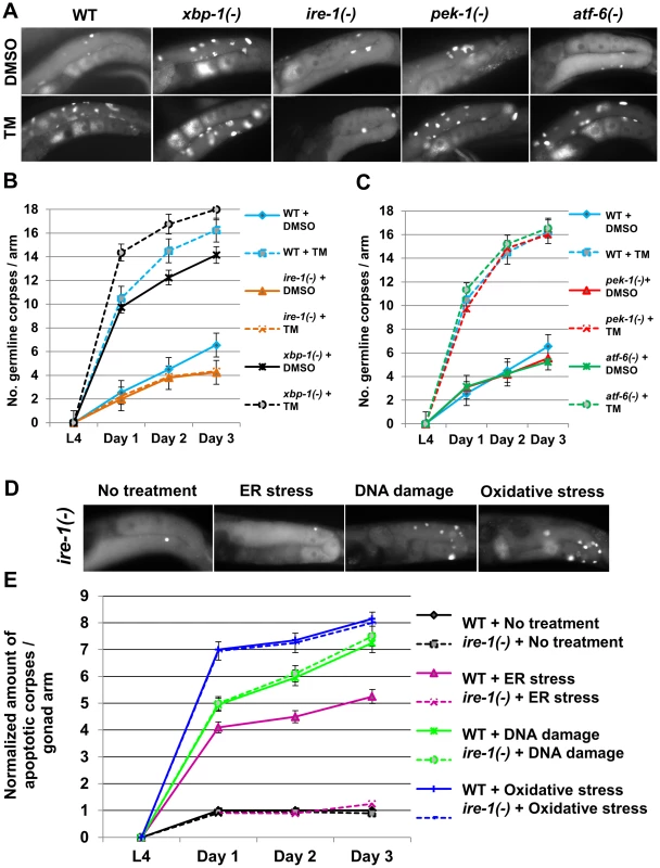
(A–C) Animals of the indicated genotypes were treated with DMSO (solid lines) or with 25 µg/ml tunicamycin (TM, dashed lines) from day-0 (L4). Germ cell corpses were identified by SYTO12 staining. (A) Representative fluorescence micrographs (400-fold magnification) of SYTO12-stained germ cell corpses of day-2 animals. (B–C) Time course experiment presenting average number of apoptotic corpses per gonad arm, from day-0 (L4) to day-3 of adulthood, 160 animals were analyzed per genotype. Error bars mark SEM. (D–E) Wild-type (WT) or ire-1(−) animals were treated with control RNAi, tfg-1 RNAi (which induces ER stress by abrogating protein export from the ER), rad-51 RNAi (to induce DNA damage) or with 10 mM paraquat (to induce oxidative stress). (D) Representative fluorescence micrographs (400-fold magnification) of SYTO12-stained germ cell corpses of control or control or stressed ire-1(−) day-2 mutants. (E) The normalized amount of apoptotic corpses per gonad arm from day-0 (L4) to day-3 of adulthood is presented. The average amount of SYTO12-labeled apoptotic corpses per gonad arm was normalized to the average number of mitotic germ cells in each of the indicated genotypes. 120 animals per treatment were analyzed. Error bars mark SEM. If indeed the increased number of germline corpses in tunicamycin-treated animals is a consequence of ER stress, then additional manipulations that disrupt ER homeostasis should also increase germ cell apoptosis. tfg-1 encodes a protein that directly interacts with SEC-16 to control COPII subunit accumulation at ER exit sites and is required for the vesicular export of cargo from the ER [34]. We hypothesized that ER homeostasis would be disrupted in tfg-1-deficient animals. To examine the effect of tfg-1 deficiency on ER homeostasis, we assessed the effect of tfg-1 RNAi treatment on the levels of the ER stress response reporter Phsp-4::gfp [22]. tfg-1 RNAi efficacy was confirmed by the reduction in the animals' body size compared to control RNAi treated animals [35]. We found that treatment with tfg-1 RNAi specifically activated the ER stress response, as it increased the level of the ER stress response reporter without increasing the expression of oxidative stress response, heat shock response or mitochondrial stress response reporters (Figure S1).
In terms of germ cell apoptosis, we observed that tfg-1 RNAi consistently increased the number of apoptotic germ cells in the gonad by approximately 4 fold from day-1 to day-3 of adulthood compared to wild-type animals (P<0.001, Figure 2A,B). A similar 4 fold increase in germ cell apoptosis was observed by scoring germ cell engulfment by neighboring cells that expressed GFP-labeled CED-1, a transmembrane receptor that mediates cell corpse engulfment in C. elegans [36] (Figure 2C). tfg-1 RNAi treatment did not increase the number of SYTO12-labeled cells in the gonads of apoptosis-defective ced-3(n1286) mutants, confirming that the dye specifically labels apoptotic cells (Figure S2). Together, these results indicate that conditions that disrupt ER homeostasis, including tunicamycin treatment or blocking secretory traffic from the ER, increase apoptosis frequency in the gonad compared to non-stressed animals.
Fig. 2. Genetically-induced ER stress increases germ cell apoptosis in an ire-1-dependent manner. 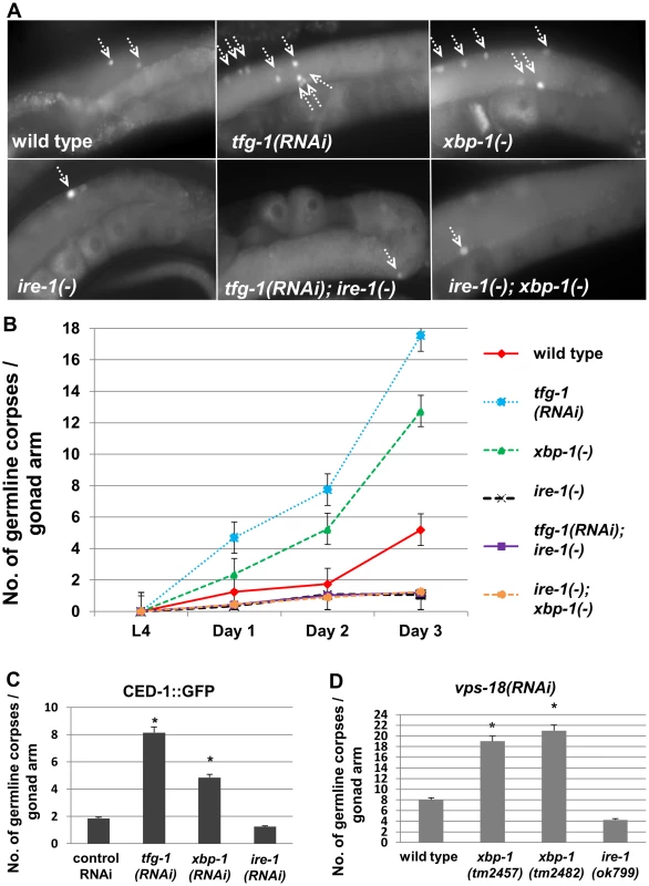
(A–B) Animals of the indicated genotypes were treated with control or tfg-1 RNAi. Germ cell corpses were identified by SYTO12 staining. (A) Representative fluorescence micrographs (400-fold magnification) of SYTO12-stained germ cell corpses in day-2 adults. Top panels present gonads of wild-type, xbp-1(tm2457) or tfg-1 RNAi treated animals. Bottom panels present gonads of the corresponding genotypes in an ire-1(ok799) background. Arrows point at SYTO12-stained germ cell corpses. (B) Time course analysis of cell corpses in the adult gonads of the indicated genotypes. SYTO12-labeled cell corpses were scored at L4, day 1, day 2 and day 3 of adulthood. Each point represents the mean number of cell corpses scored in 30 gonads. Error bars indicate SEM. (C) Animals expressing CED-1::GFP were treated with control, tfg-1, xbp-1 or ire-1 RNAi and analyzed. Bar graph shows average number of apoptotic corpses per gonad arm in the indicated genotypes identified by their engulfment by CED-1::GFP labeled gonadal sheath cells. Asterisk indicates a significant increase in germline apoptosis compared to control RNAi-treated animals (Student's t-test values of P<0.001). (D) Bar graph shows average number of SYTO12-labeled apoptotic corpses per gonad arm in day-2 vps-18 RNAi-treated animals (n = 40 gonads per genotype). Asterisk indicates a significant increase in germline apoptosis compared to wild-type animals treated with vps-18 RNAi) Student's t-test values of P<0.001). ER stress-induced germ cell apoptosis is mediated by the apoptotic machinery implicated in DNA damage-induced germ cell apoptosis
We next asked which apoptotic machinery is implicated in ER stress-induced germ cell apoptosis. To this end, we examined mutants deficient in core-apoptotic genes as well as mutants deficient in genes specifically implicated in germ cell apoptosis. This array of apoptosis-related mutants was treated with control or tfg-1 RNAi, and germ cell apoptosis was scored by SYTO12 labeling. As expected, we found that the core apoptosis machinery genes ced-3 and ced-4 [6], [13], [15], [16] were required for germ cell apoptosis in response to ER stress (Figure 3A). Importantly, the cep-1, egl-1 and ced-13 genes, previously implicated in DNA damage-induced apoptosis [9], [17]–[19], were also found to be completely essential for germ cell apoptosis in response to tfg-1 RNAi treatment (Figure 3A). Accordingly, the levels of a CEP-1::GFP translational fusion transgene driven by the cep-1 promoter were increased within the germ cells of tfg-1 RNAi-treated animals (P<0.001, Figure 3B). In contrast, pmk-1 and sek-1, previously implicated in oxidative stress-induced and pathogen-induced germ cell apoptosis respectively [7], [8], were dispensable for germ cell apoptosis in response to tfg-1 RNAi treatment (Figure 3C). Thus, the genetic analysis clearly implicated the apoptotic machinery that mediates DNA damage-induced germ cell apoptosis in ER stress-induced germ cell apoptosis as well.
Fig. 3. ER stress-induced germ cell apoptosis is mediated by the same apoptotic machinery implicated in DNA damage-induced apoptosis. 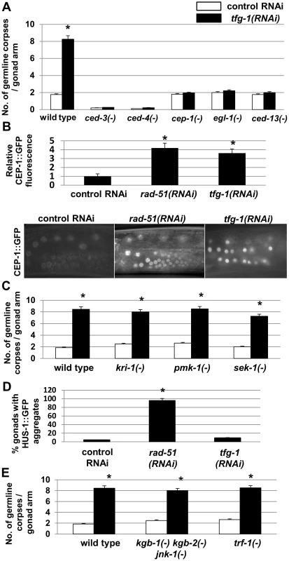
(A,C,E) Bar graphs present average number +/−SEM of SYTO12-labeled apoptotic corpses per gonad arm in day-2 animals of the indicated genotypes treated with control RNAi (white bars) or tfg-1 RNAi (black bars). At least 50 gonads per genotype were scored. Asterisks mark Student's t-test values of P<0.001 between tfg-1 RNAi and control RNAi. (B) Mean fluorescence and representative fluorescence micrographs (400-fold magnification) of CEP-1::GFP expression in the germ cells of CEP-1::GFP transgenic animals treated with control, rad-51 or tfg-1 RNAi. Each bar graph presents the average fluorescence of 100 germ cells from 10 gonads. rad-51 RNAi treatment served as a positive control for DNA damage-induced expression of CEP-1::GFP. Asterisks mark Student's t-test values of P<0.001 compared to control RNAi treatment. (D) Bar graph presents percentage of dissected gonads of HUS-1::GFP transgenic animals treated with control, rad-51 or tfg-1 RNAi, in which HUS-1::GFP aggregates were detected. 50–60 transgenic gonads were analyzed. rad-51 RNAi treatment served as a positive control for DNA damage-induced HUS-1::GFP aggregation. Asterisks mark Student's t-test values of P<0.001 compared to control RNAi treatment. Error bars present SEM. The strong changes in CEP-1 levels observed in tfg-1 RNAi-treated animals suggest that ER stress controls CEP-1 activation within the germ cells. One possible explanation for the involvement of genes implicated in DNA damage-induced germ cell apoptosis is that ER stress indirectly damages DNA, which in turn leads to CEP-1 activation and DNA damage-induced germ cell apoptosis. However, whereas a previous study implicated the intestinal kri-1 gene in non-autonomous regulation of ionizing-radiation induced germ cell apoptosis [37], we found that tfg-1 RNAi treatment efficiently induced germ cell apoptosis in kri-1-deficient animals (Figure 3C). This genetic uncoupling between the requirements for ionizing-radiation induced germ cell apoptosis and ER stress-induced germ cell apoptosis suggest that ER stress does not simply induce DNA damage which in turn leads to germ cell apoptosis.
To further substantiate this conclusion, we examined directly whether the DNA damage response is activated in the germ cells of ER stressed - animals treated with tfg-1 RNAi. To this end, we followed the nuclear aggregation of HUS-1::GFP, which encodes a DNA damage checkpoint protein that relocalizes to distinct nuclear foci upon induction of DNA damage [38]. Whereas nuclear HUS-1::GFP aggregates were clearly observed in the germ cells of DNA-damaged rad-51 RNAi treated animals (P<0.001 compared to control RNAi), HUS-1::GFP aggregates were not detected in tfg-1 RNAi treated animals (P = 0.78 compared to control RNAi, Figure 3D). Thus, ER stress activates CEP-1/p53 to induce germ cell apoptosis without generating DNA damage.
Conditions that disrupt ER homeostasis induce germ cell apoptosis via the ER stress sensor ire-1
We next asked whether any of the canonical ER stress sensing genes was implicated in ER stress-induced germ cell apoptosis. To this end, we examined mutants deficient in the ER stress-response sensor genes that comprise the UPR: ire-1, pek-1 or atf-6. This array of mutants was treated with DMSO or with tunicamycin and germ cell apoptosis was scored by SYTO12 labeling. We found that similarly to its effect in wild-type animals, tunicamycin treatment increased the number of germline corpses in atf-6 and pek-1-deficient animals by approximately 3 fold from day-1 to day-3 of adulthood (P<0.001, Figure 1A,C). In contrast, ER stress induced by tunicamycin treatment failed to increase germ cell apoptosis in ire-1 mutants (Figure 1A,B). Therefore, the insensitivity of the germline to tunicamycin is unique to ire-1-deficient animals and not seen in animals deficient in other UPR sensors.
ire-1 mutants are abnormal in terms of their gonad anatomy and their reproductive capacity: ire-1 mutants have approximately 2 fold less progeny and 2 fold less mitotic germ cells within their proliferative zones compared to ire-1(+) wild-type animals (P<0.001, Figures S3A,D). Thus, we wondered whether these abnormalities affected the ability of their germ cells to undergo apoptosis in general or whether they were specifically defective in their ability to undergo apoptosis in response to ER stress.
First we assessed germline apoptosis in ire-1(−) mutants under normal growth conditions, from day-0 (L4) to day-3 of adulthood. At all timepoints, we detected approximately half the amount of germline corpses in ire-1(−) gonads compared to ire-1(+) wild-type gonads, as assessed by SYTO12 labeling and by the CED-1::GFP engulfment marker (Figure 2A–C). The low levels of germ cell apoptosis persisted in ire-1 mutants treated with vps-18 RNAi (Figure 2D), a treatment that impairs germ cell corpse clearance [39]. However, normalization of the number of apoptotic corpses to the number of mitotic germ cells resulted in comparable levels of germ cell corpses in ire-1 mutants and in non-stressed wild-type animals (P = 0.12, Figure S3C). This indicates that in spite of their reproductive abnormalities, physiological germ cell apoptosis in ire-1(−) and ire-1(+) animals is comparable.
Next, we assessed stress-induced germline apoptosis in ire-1 mutants. We found that although manipulations that disrupt ER homeostasis fail to increase germ cell apoptosis in ire-1 mutants (Figure 1B,1D–E), DNA damage and oxidative stress conditions did increase germ cell apoptosis in ire-1 mutants (P<0.001 compared to non-stressed ire-1 mutants, Figure 1D–E). This resulted in a similar level of germline apoptosis as in stressed wild-type animals upon normalization of the number of apoptotic corpses to the number of mitotic germ cells (P = 0.44 for DNA damage and P = 0.42 for oxidative stress, Figure 1D–E). Thus, in spite of the reproductive abnormalities of ire-1 mutants, the germ cells of these mutants undergo stress-induced apoptosis similarly to wild-type animals, however not in response to ER stress. The inability of ire-1 mutants to increase germline apoptosis specifically in response to perturbations in ER homeostasis suggests that IRE-1 may be a critical mediator of ER stress-induced germ cell apoptosis.
IRE-1 function in ER stress-induced germ cell apoptosis is independent of its canonical downstream target XBP-1
The most established mode of action of IRE-1 under ER stress conditions is via the activation of the UPR-related transcription factor XBP-1 [22]–[24]. Therefore, if IRE-1 enabled ER stress-induced germ cell apoptosis via its downstream target xbp-1, then the number of germline corpses detected in xbp-1(−) mutants should remain low under ER stress conditions, similarly to ire-1(−) mutants.
In order to test this, we first examined germ cell apoptosis in xbp-1(tm2457) null mutants. Surprisingly, in contrast to ire-1(−) mutants, we consistently detected increased germ cell apoptosis in xbp-1(−) gonads compared to wild-type gonads under normal growth conditions. A 2.5 fold increase in the number of germline corpses in xbp-1(−) mutants was detected by SYTO12 labeling of gonads from day-1 to day-3 of adulthood compared to wild-type animals (P<0.001, Figure 2A–B). A similar observation was apparent by using the CED-1::GFP engulfment marker (Figure 2C). The 2.5 fold increase in the number of germ cell corpses was still apparent in engulfment defective vps-18 RNAi-treated animals (Figure 2D) and upon normalization to the number of mitotic germ cells located in the proliferative zone (Figure S3C). Thus, in contrast to ire-1 mutants and wild-type animals, xbp-1 mutants exhibit a high basal level of germ cell apoptosis.
We hypothesized that the increase in germline apoptosis in xbp-1 mutants may be due to perturbed ER homeostasis in these animals [25], [26], If so, then it should be mediated via ire-1, similarly to other ER stress conditions that induce germ cell apoptosis. Accordingly, we found that in an ire-1(−) background, the xbp-1 mutation did not increase germ cell apoptosis. This observation was consistent along different time points spanning from day-0 (L4) to day-3 of adulthood (Figure 2A–B). This also persisted upon normalization to the number of mitotic germ cells located in the proliferative zone (Figure S3C). Interestingly, the amount of mitotic germ cells of xbp-1; ire-1 double mutants was similar to that of xbp-1 single mutants (P = 0.072, Figure S3A), whose germ cells were responsive to ER stress-induced apoptosis (Figure S3B–C). This indicates that the reduced amount of mitotic cells in the gonad of ire-1 mutants can be uncoupled from the inability of their germ cells to undergo ER stress-associated apoptosis.
The finding that xbp-1 deficiency per se promotes ire-1-dependent ER stress-induced germ cell apoptosis suggests that xbp-1 is dispensable for increasing germ cell apoptosis in response to ER stress. Consistent with this, we found that tunicamycin treatment further increased germ cell apoptosis in xbp-1 mutants (P<0.001, Figure 1A–B). Altogether, these results lend further support to the notion that ire-1 is a critical signaling molecule in mediating ER stress-induced germline apoptosis, whereas its' downstream canonical target xbp-1 is not. Furthermore, since ER function is compromised both in ire-1 and in xbp-1 deficient mutants [25], [26], the differential ability to induce germ cell apoptosis in these mutants suggests that germ cell apoptosis may be the result of active IRE-1 signaling, rather than simply a consequence of ER dysfunction.
In mammalian cells, activation of IRE1 can cell-autonomously activate JNK via the adaptor protein TRAF. Consequently, IRE1-mediated activation of JNK initiates proapoptotic signaling, independently of XBP1 [29]. Thus, we examined whether the C. elegans homologs of TRAF and JNK proteins were required for ire-1/ER stress-induced apoptosis in C. elegans, which is also independent of xbp-1. To this end, trf-1 mutants or mutants deficient in all three C. elegans JNK homologs were treated with control or tfg-1 RNAi. We found that tfg-1 RNAi increased germ cell apoptosis independently of the trf-1 and the JNK-like genes (Figure 3E). Thus, since ER stress can effectively induce germ cell apoptosis in the absence of xbp-1, trf-1 and JNK homologs, the signaling mediated by IRE-1, in this case, must be executed by an alternative xbp-1-independent output of IRE-1.
ER stress in the ASI sensory neurons regulates germ cell apoptosis cell non-autonomously
Next, we examined whether ER stress triggers programmed cell death autonomously within the germ cells, or non-autonomously from the soma. To test this, we used tfg-1 RNAi to induce ER stress specifically in the germline or in the soma. To induce ER stress primarily in the germ cells, mutants in the rrf-1 gene, encoding an RNA-directed RNA polymerase (RdRP) homolog required for most somatic RNAi but not for germline RNAi [40], were treated with tfg-1 RNAi. No increase in the amount of germline corpses was observed as a result of tfg-1 RNAi treatment in rrf-1 mutants (P = 0.19, Figure 4A). To induce ER stress specifically in the soma, mutants in the ppw-1 gene, which is required for efficient RNAi in the germline [41], were treated with tfg-1 RNAi. This resulted in a 4.5 fold increase in the amount of apoptotic corpses in the gonads (P<0.001, Figure 4A). Thus, ER stress in the soma, rather than in the germ cells, is sufficient for the induction of germ cell apoptosis.
Fig. 4. ER stress specifically in sensory neurons is sufficient to induce germline apoptosis. 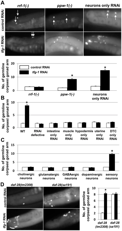
(A) Bar graph and representative fluorescence micrographs (400-fold magnification) of SYTO12-stained germ cell corpses in day-2 adults. Arrows point at SYTO12 stained germ cell corpses. Bar graph presents average number +/−SEM of apoptotic corpses per gonad arm in the indicated genotypes (n = 60 per genotype). Asterisks mark Student's t-test values of P<0.001 of tfg-1 RNAi treated animals compared to the corresponding control RNAi treated animals. Note that in animals in which RNAi functions only in the neurons, tfg-1 RNAi increased the amount of germ cell corpses to a greater extent than in ppw-1 mutants or wild-type animals. This may be due to more efficient tfg-1 RNAi uptake in the neurons of these animals, whose neurons over-express SID-1 [76] (B,C) Bar graph presents average number +/− SEM of SYTO-12-labeled germ cell corpses of day-2 adults of the indicated genotypes (n = 60 gonads per genotype). Asterisks mark Student's t-test values of P<0.001 of tfg-1 RNAi treated animals (black bars) compared to the corresponding control RNAi treated animals (white bars). See materials and methods for strains details. (D) Representative fluorescence micrographs (400-fold magnification) and bar graph of SYTO12-stained germ cell corpses in day-2 daf-28 mutant strains treated with control RNAi (white bars) or tfg-1 RNAi (black bars). Bar graph presents average number +/− SEM of apoptotic corpses per gonad arm in the indicated genotypes (n = 70 per genotype). tm2308 is a deletion mutation in the daf-28 gene. sa191 is a point mutation that interferes with the posttranslational processing of DAF-28. Asterisks mark Student's t-test values of P<0.001 of tfg-1 RNAi treated animals compared control RNAi treated animals. Does germ cell apoptosis occur upon disruption of ER homeostasis in the entire soma or does it occur in response to ER stress in a particular part of the soma? To answer this, ER stress was induced locally in specific somatic tissues. This was achieved by treating animals expressing functional RNAi machinery only in specific tissues with tfg-1 RNAi and assessing germ cell apoptosis in these animals. We found that tfg-1 RNAi treatment did not increase germ cell apoptosis in animals which respond to RNAi only in the intestine, in the muscle, in the hypodermis, in the uterine or in the distal tip cells (P>0.1 in each one of these strains, Figure 4B). In contrast, tfg-1 RNAi treatment increased germ cell apoptosis by approximately 7 fold in animals which respond to RNAi specifically in the neurons (P<0.001, Figure 4A).
Next, we examined whether ER stress-induced germline apoptosis is under pan-neuronal control or under the control of specific neurons. To this end, we introduced ER stress-inducing tfg-1 RNAi into animals expressing functional RNAi machinery specifically in the cholinergic, glutamatergic, GABAergic, dopaminergic or in a subset of sensory neurons. Importantly, we found that tfg-1 RNAi treatment increased germ cell apoptosis only in animals whose sensory neurons responded to RNAi (Figure 4C).
Among the sensory neurons whose exposure to ER stress increased germline apoptosis were the ASI neurons, which have been previously implicated in the regulation of germ cell proliferation and maturation [42]. Hence, we examined whether ER stress in the ASI sensory neurons alone is sufficient for the induction of germ cell apoptosis in the gonad. To this end, we first assessed germline apoptosis in daf-28(sa191) mutants, which produce a toxic insulin peptide that activates the UPR specifically in the ASI neurons [43]. We found that germ cell apoptosis in the gonads of daf-28(sa191) mutants was increased by approximately 4 fold compared to wild-type animals (P<0.001, Figure 4D). Importantly, germ cell apoptosis was not increased in a daf-28(tm2308) null strain, which is deficient in daf-28 and does not produce the toxic insulin peptide which induces ER stress (P = 0.15, Figure 4D). tfg-1 RNAi treatment of the two daf-28 mutant strains increased the number of germline corpses in daf-28(tm2308) null strain (P<0.001), but did not further increase germline apoptosis in the daf-28(sa191) strain (P = 0.09, Figure 4D). tfg-1 RNAi treatment did not alter ASI overall morphology as assessed by the expression pattern of a GFP reporter driven by an ASI-specific promoter (Figure S4A). Together, these findings suggest that expression of the toxic form of DAF-28 and tfg-1-deficiency increase germ cell apoptosis by similar means; most likely by causing ER stress and activating the UPR in the ASI neurons.
Activation of IRE-1 in ASI neurons is sufficient to induce germ cell apoptosis
We have demonstrated that in the absence of the ER stress sensor ire-1, ER stress does not increase germline apoptosis. We further demonstrated that ER stress in the ASI sensory neurons is sufficient to induce germ cell apoptosis. Thus, we next examined whether it is also sufficient to express ire-1 in the soma, and specifically in the ASI neurons, to restore germ cell apoptosis in response to ER stress.
To this end, we restored ire-1 expression in the entire soma, pan-neuronally or specifically in the ASI/ASJ neurons of ire-1(−) mutants. This was achieved using multi-copy ire-1 transgenes under ire-1, rgef-1 and daf-28 promoters respectively. Since the expression of multi-copy transgenes is normally suppressed in germ cells [44], and due to the specificity of their promoters, these transgenes restore ire-1 expression within different parts of the soma but not in the germline. We found that expression of each of these ire-1 transgenes completely restored the increase in germline apoptosis in response to treatment with tfg-1 RNAi (P<0.001, compare white and black bars within each strain in Figure 5A). Similarly, we restored ire-1 expression in muscle cells and in the PVD and OLL neurons using multi-copy ire-1 transgenes under myo-3 and ser-2 promoters respectively. No increase in germline apoptosis in response to tfg-1 RNAi treatment was apparent in these two transgenic lines compared to control RNAi treatment (P>0.1, Figure 5A). The fact that not all ire-1 transgenes induced apoptosis supports the notion that ire-1-induced germline apoptosis is not the result of leaky expression of the transgenes in other tissues. Altogether, this implies that not all tissues and not all neurons are involved in the regulation of this process.
Fig. 5. Over-expression of ire-1 in the ASI pair of amphid neurons is sufficient to induce germline apoptosis independently of ER stress. 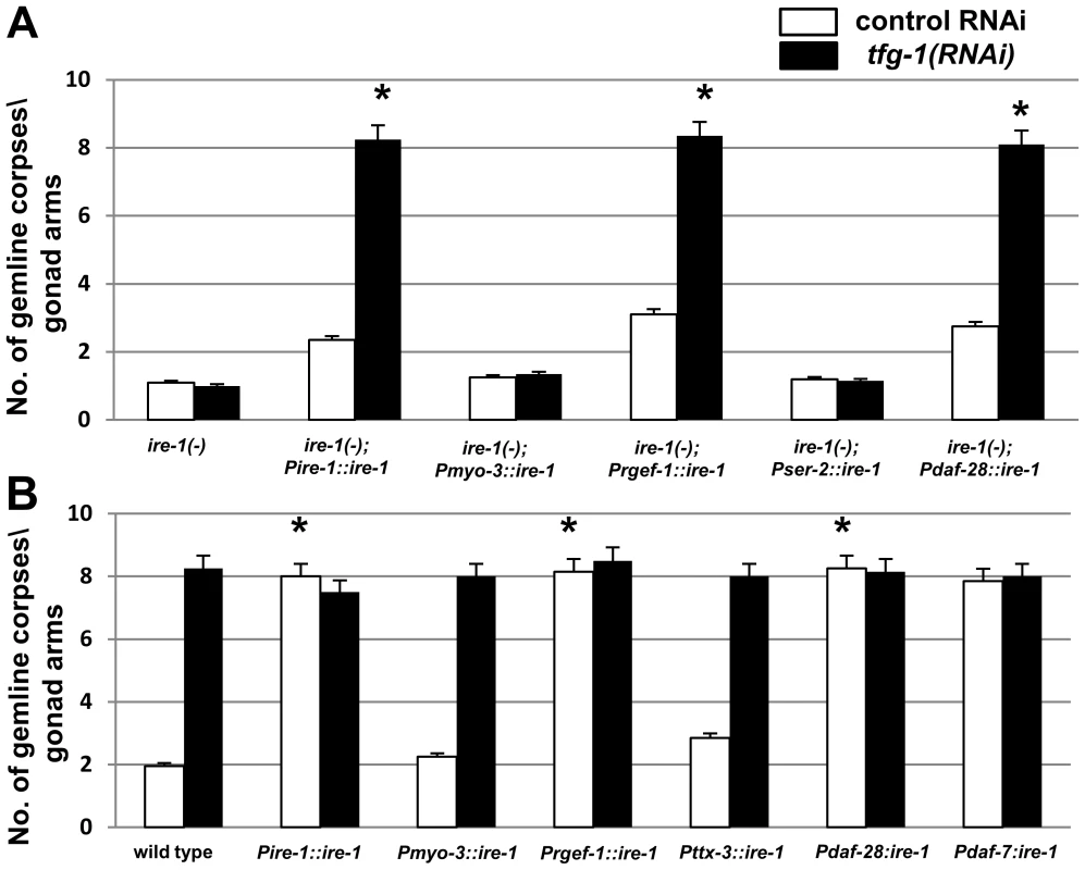
(A–B) Bar graph presents average number of apoptotic corpses per gonad arm in day-2 ire-1(−) or ire-1(+) animals expressing an ire-1 transgene driven by the indicated promoters (n = 100 gonads per genotype). Animals were treated with control RNAi (white bars) or with tfg-1 RNAi (black bars). Asterisks mark Student's t-test values of P<0.001 of each strain treated with tfg-1 RNAi compared to its' control RNAi treatment. Error bars indicate SEM. (A) Germ cell apoptosis in ire-1(−) mutants expressing an ire-1 rescuing transgene in different tissues is presented. The ire-1 rescuing transgene was expressed in somatic cells (Pire-1::ire-1), in neurons (Prgef-1::ire-1, Pdaf-28::ire-1 and Pser-2::ire-1, which drive expression in all neurons, in the ASI/ASJ neurons or in the PVD and OLL neurons respectively) and in muscle cells (Pmyo-3::ire-1). (B) Germ cell apoptosis in ire-1(+) animals overexpressing an ire-1 transgene in different tissues is presented. The ire-1 transgene was expressed in somatic cells (Pire-1::ire-1), in neurons [driven by the promoters of rgef-1 (pan-neuronal expression), daf-28 (expressed in the ASI/ASJ neurons), daf-7 (expressed only in the ASI neurons) and ttx-3 (expressed in the AIY interneurons)] and in muscle (Pmyo-3::ire-1). Next, we asked whether increasing IRE-1 levels to a greater extent may be sufficient for inducing germline apoptosis even in the absence of ER stress. To this end, we overexpressed ire-1 transgenes in various tissues or cells of ire-1(+) wild-type animals. This was achieved by using multi-copy ire-1 transgenes under ire-1, rgef-1, daf-28 and daf-7 promoters. This is consistent with the interpretation that some activation of IRE-1 is achieved merely by its over-expression, as has been previously observed in yeast and in mammalian cells [45], [46]. We found that this artificial activation of IRE-1 in the soma, pan-neuronally or specifically in the ASI/ASJ neurons of ire-1(+) animals was sufficient to induce high levels of germ cell apoptosis (P<0.001, compare white bars of transgenic animals to that of wild-type animals in Figure 5B). No increase in germ-cell apoptosis was observed upon overexpression of an ire-1 transgene in muscle cells or in the AIY neurons in ire-1(+) animals (P>0.1, Figure 5B). These findings support the claim that the rescuing activity of the ire-1 transgenes stems from their expression in specific neurons.
tfg-1 RNAi treatment of ire-1(+) animals over-expressing the ire-1 transgenes in the soma, in the neurons and specifically in the ASI/ASJ neurons did not further increase germline apoptosis (P>0.5, compare white and black bars within the strains, Figure 5B). This suggests that IRE-1 overexpression and tfg-1-deficiency increase germ cell apoptosis by similar means, i.e. by activating IRE-1. Taken together, our data demonstrate that activation of ire-1 specifically in the ASI neurons, either by ER stress in the ASI neurons or by IRE-1 overexpression, can non-autonomously regulate germ cell apoptosis. Furthermore, since over-expression of transgenic IRE-1 is sufficient for its artificial activation in a manner that is independent of ER stress, this further suggests that active IRE-1 signaling in the ASI neurons per se, rather than neuronal ER stress or ER dysfunction, is the cause of germ cell apoptosis.
Discussion
Understanding the molecular events that regulate the life-death decision of cells is of fundamental importance in cell biology research, cell development, cancer biology and disease biology [47]. In this study, we gained new and fascinating insights into the complex coupling between ER stress in the nerve system and germ cell apoptosis.
We report for the first time that germ cells undergo apoptosis in response to ER stress. We find that activation of the ER stress response gene ire-1 is required and sufficient to induce germ cell apoptosis in response to several ER stress-inducing conditions. Strikingly, we find that germ cell fate is regulated non-autonomously by ER stress and/or through IRE-1 activation specifically in the ASI neurons. This implies that ER homeostasis and UPR signaling in the germ cells themselves is not a factor in determining their fate, ruling out the possibility that these apoptotic events are part of a quality control process that removes “stress-damaged” germ cells from the gonad [6], [11], [12]. Furthermore, this assigns a central neuroendocrine role for the ASI neuron pair in coupling between stress sensing and the onset of germ cell apoptosis. This is in addition to other central physiological processes in C. elegans, such as dauer formation [48], [49] and longevity [50], [51], that are also controlled by the sensory ASI neuron pair. Interestingly, another pair of sensory neurons, the ASJ neurons, has been previously implicated in the protection of germ cells from apoptosis under hypoxic conditions [52]. Thus, depending on the stress condition, different neurons can shift germ cell fate from survival to death or vice versa.
How might IRE-1 activation in the ASI neurons dictate germ cell survival or death? One possibility is that defects associated with ire-1 deficiency and/or ire-1 activation indirectly abrogate the communication between the neurons and the gonad. However, several lines of evidence undermine this hypothesis: (1) We find that ER stress-induced germ cell apoptosis proceeds normally in animals with a severely defective nervous system (Figure S4B,C). This implies that germline apoptosis does not result from a generic neuronal defect. (2) ire-1 deficiency is associated with germline abnormalities which include a significant reduction in the number of mitotic germ cells and in reduced progeny number. However, these gonad-related defects do not confer generic resistance to stress-induced apoptosis as the germ cells of ire-1 mutants do undergo apoptosis in response to a variety of stresses. Furthermore, a mutation in xbp-1, which improved the reproductive abnormalities of ire-1 mutants, did not restore responsiveness to ER stress induced germ cell apoptosis in ire-1; xbp-1 double mutants, thus uncoupling the two. (3) Whereas the comparison of germ cell apoptosis in ire-1 and wild-type animals may be confined by the basal discrepancy of their reproductive systems, this concern does not exist in the analysis of ire-1 overexpressing strains, whose gonad appears to be normal (P>0.1 for Pire-1::ire-1 and Pdaf-7::ire-1 compared to wild-type animals Figure S3A,D). Similarly, this concern does not exist in the intra-strain comparisons of germline apoptosis within the ire-1(−) strain under control and stress conditions.
If ire-1 misregulation in the ASI neurons does not indirectly abrogate the communication between the neurons and the gonad, how might it dictate germ cell survival or death? IRE-1 is a dual-activity enzyme, bearing both kinase and endoribonuclease activities and a propensity to self-aggregate at the ER membrane in response to ER stress. The most characterized mode of action of IRE-1 is the activation of its downstream transcription factor XBP-1 [53]. Significantly less characterized are XBP-1 independent targets of IRE-1, that include activation of the cell death machinery via JNK/TRAF signaling and degradation of ER-localized mRNAs that encode secreted and membrane proteins in a process called RIDD [29], [30], [32], [54]–[58]. Since we find that ER stress can effectively induce germ cell apoptosis in the absence of xbp-1, trf-1 and JNK homologs, the signaling mediated by IRE-1 in this case may be executed by the RIDD pathway or via a novel, yet undescribed, xbp-1-independent output of IRE-1.
We propose that activation of IRE-1 in the neurons (either as a result of ER stress or merely by its over-expression) actively regulates the production of a germ cell regulatory signal. In principle this may be a germ cell proapoptotic signal produced by the neurons upon IRE-1 activation. Alternatively, this may be a germ cell anti-apoptotic signal that is down-regulated by IRE-1 upon its activation. This ASI-regulated signal, whose identity and nature remain to be elucidated, propagates in the animal and affects the gonad where it acts upstream to the p53 homolog cep-1, activating the same apoptotic machinery in the germ cells as the DNA damage response, without inducing DNA damage in the germ cells (Figure 6). This indicates the existence of a new pathway that can activate CEP-1 independently of DNA damage upon activation of neuronal IRE-1.
Fig. 6. Model – Activation of IRE-1 in the ASI neurons induces germ cell apoptosis. 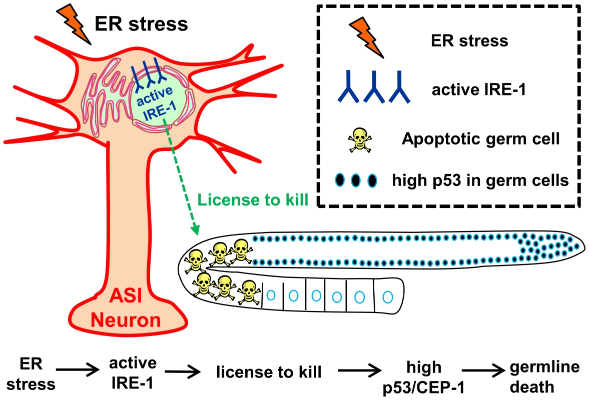
Our data demonstrate that activation of IRE-1 in ASI neurons (by ER stress or simply by its overexpression) is sufficient and required for ER stress-related induction of germline apoptosis. We propose that activation of IRE-1 in the ASI neurons initiates a signaling cascade that “licenses” germ cell apoptosis. This, in turn, increases CEP-1 levels in the germ cells, which triggers germ cell apoptosis using the same genes previously associated with DNA damage induced apoptosis. Interestingly, in adult animals, exposure to ER stress or activation of IRE-1 in the soma induce apoptosis only in germ cells, as we did not detect any apoptotic corpses outside of the gonad of these animals. This is in contrast to the developing embryo, where exposure to ER stress can induce apoptosis in the soma [35], [59]. We propose that as the organism completes development, its ability to respond or execute programmed cell death upon exposure to ER stress is maintained in mitotic germ cells while being selectively abrogated in the post-mitotic soma, as has been demonstrated for their ability to execute apoptosis in response to DNA-damage [60]. This resistance of the soma is important in terms of survival of the animal as it prevents cell death of somatic tissues that lack stem cell pools and regenerative capacity, while allowing cell death of immortal germline cells at times of stress.
What could be the advantage in diluting the germ cell pool when neurons “feel” ER-stressed (i.e. when IRE-1 is activated naturally by ER stress or artificially by overexpression)? Recent studies demonstrate a tight inverse correlation between germ-cell proliferation and the maintenance of somatic proteostasis and longevity [61]–[65]. This inverse correlation is thought to be due to a limitation of resources shared by the germline and the soma and due to altered metabolic and cellular repair mechanisms in the soma that are enabled upon germ cell loss. Previous studies implicated the nervous system in systemic and hierarchical control of cellular stress responses elsewhere in the soma to maintain organismal homeostasis [66]–[72]. Our data further imply that neurons also have the ability to communicate with the germ cells to promote their death in response to stress in the ER. This, in turn, may orchestrate a proteostasis switch in the soma at the expense of a replenishable germ cell pool in times of stress. This adds a new layer of complexity to our understanding of how protein homeostasis is regulated and coordinated across tissues in multicellular organisms.
Materials and Methods
Cell corpse assay
For single time-point experiments, the number of apoptotic germ cells was scored in day-2 animals stained with SYTO12 (Molecular Probes) as previously described [6]. For time course experiments, the number of SYTO12-labeled apoptotic corpses per gonad arm was scored in animals from day-0 (L4) to day-3 of adulthood. Where indicated, the average number of apoptotic corpses was normalized to the number of mitotic germ cells within the proliferative zone of the gonads, determined by section analysis of DAPI-stained gonads.
Oxidative stress treatment
Day-1 adult animals were placed in 200 µl of M9 (control) or 10 mM paraquat (oxidative stress) for 1.5 h at 20°C. After the incubation period, 1 ml of M9 was added to dilute the paraquat. Animals were then transferred to eppendorfs with SYTO12 staining for 4.5 hrs. Animals were allowed to recover on plates for 40 min. Finally, the animals were mounted and observed under the microscope to determine cell corpse numbers.
Gonad dissection and DAPI staining
Gonads of day-1 adults were dissected, fixed, and stained with DAPI as previously described [10].
RNA interference
Bacteria expressing dsRNA were cultured overnight in LB containing tetracycline and ampicillin. Bacteria were seeded on NGM plates containing IPTG and carbenicillin. RNAi clone identity was verified by sequencing. Eggs were placed on plates and synchronized from day-0 (L4).The efficacy of the tfg-1 RNAi was confirmed by the animals' reduced body size [35].
Fluorescence microscopy and quantification
Animals were anaesthetized on 2% agarose pads containing 2 mM levamisol.
Images were taken with a CCD digital camera using a Nikon 90i fluorescence microscope. For each trial, exposure time was calibrated to minimize the number of saturated pixels and was kept constant through the experiment. The NIS element software was used to quantify mean fluorescence intensity as measured by intensity of each pixel in the selected area.
Statistical analysis
Error bars represent the standard error of the mean (SEM) of at least 3 independent experiments. P values were calculated using the unpaired Student's t test.
Strains and transgenic lines
The following lines were used in this study: N2, CF2012: pek-1(ok275) X, CF2988: atf-6(ok551) X, CF2473: ire-1(ok799) II, CF3208: xbp-1(tm2457) III, SHK62: ire-1(ok799) II; xbp-1(tm2457) III, MD701: Plim-7 ced-1::gfp V, xbp-1(tm2482) III, CF2185: ced-3(n1289) IV, MT2547: ced-4(n1162) III, TJ1: cep-1(gk138) I, MT8735: egl-1(n1084n3082) V, FX536: ced-13(tm536) X, WS1433: hus-1(op241) I; unc-119(ed3) III; opIs34, CF2052: kri-1(ok1251) I, KU25: pmk-1(km25) IV, AU1: sek-1(ag1) X, CF3030: kgb-1(um3) kgb-2(gk361) jnk-1(gk7) IV, NS2937: trf-1(nr2014) III, CF2260: zcIs4[Phsp-4::gfp] V, CL2166: Pgst-4::gfp(dvls19) V, CF1553: muIs84 [(pAD76) Psod-3::gfp+rol-6], SJ4100: Phsp-6::gfp(zcIs13) V, CL2070: Phsp-16.2::gfp(dvls70) V, SHK57: xbp-1(tm2457) III; ced-3(n1286) IV, SHK189: zcIs4[Phsp-4::gfp] V; Pire-1::ire-1, NL2098: rrf-1(pk1417) I, NL2550: ppw-1(pk2505) I, SHK185: ire-1(ok799) II; Prgef-1::ire-1, SHK4: Pire-1::ire-1, SHK182: Prgef-1::ire-1, BB22 rde-4(ne299) III; adr-2(gv42) III, TG12: cep-1(lg12501) I; unc-119(ed4) III; gtIs1 [CEP-1::GFP+unc-119(+)], SHK8: Pmyo-3::ire-1, SHK14: Pmyo-3::ire-1; ire-1(ok799) II, Pser-2::ire-1; ire-1(ok799) II, SHK15: Pdaf-28::ire-1; ire-1(ok799) II, SHK6: Pdaf-28::ire-1, SHK237: Pttx-3::ire-1; Pdaf-7::gfp, SHK234: Pdaf-7::ire-1; Pdaf-7::gfp, daf-28(tm2308) V, CF2638: daf-28(sa191) V, VB1605: svls69[Pdaf-28::daf-28::gfp], SHK11: ire-1(ok799) II; svls69[Pdaf-28::daf-28::gfp], SHK27: ire-1(ok799) II; Pire-1::ire-1; svls69[Pdaf-28::daf-28::gfp], SHK60: unc-13(e51) I, SK7: unc-64(e246) III; unc-31(e928) IV and MT6308: eat-4 (ky5) III.
The following strains were used for tissue-specific RNAi experiments: TU3401: sid-1(pk3321) V; Punc-119::sid-1 (neuron only RNAi), VP303: rde-1(ne213) V; Pnhx-2::rde-1 (intestine only RNAi), WM118: rde-1(ne300) V; Pmyo-3::rde-1(muscle only RNAi), NR222: rde-1(ne219) V; Plin-26::rde-1 (hypodermis only RNAi), NK640: rrf-3(pk1426)II; rde-1(ne219) V; Pfos-1A::rde-1 (uterine only RNAi),JK4143: rde-1(ne219) V; Plag-2::rde-1::gfp (distal tip cell only RNAi).
The following strains were used for neuron-specific RNAi treatments as previously described [73]: XE1581: wpSi10 II [unc-17p::rde-1::SL2::sid-1+Cbr-unc-119(+)]; eri-1(mg366) IV; rde-1(ne219) V; lin-15B(n744) X - Cholinergic neuron-specific RNAi strain. XE1375: wpIs36 I [unc-47p::mCherry]; wpSi1 II [unc-47p::rde-1::SL2::sid-1+Cbr-unc-119(+)]; eri-1(mg366) IV; rde-1(ne219) V; lin-15B(n744) X - GABAergic neuron-specific RNAi strain. XE1582: wpSi11 II [eat-4p::rde-1::SL2::sid-1+Cbr-unc-119(+)] II.; eri-1(mg366) IV; rde-1(ne219) V; lin-15B(n744) X - Glutamatergic neuron-specific RNAi strain. XE1474: wpSi6 II [dat-1p::rde-1::SL2::sid-1+Cbr-unc-119(+)] II; eri-1(mg366) IV; rde-1(ne219) V; lin-15B(n744) X - Dopaminergic neuron-specific RNAi strain, SHK231: sid-1(pk3321) V; Pche-12::sid-1(+); rol-6(su1006).
Plasmid construction
Prgef-1::ire-1 - ire-1 cDNA was cloned under the 3.5 kb rgef-1 (F25B3.3) promoter and injected at 5 ng/µl with Pmyo-3::mCherry at 50 ng/µl.
Pire-1::ire-1 - ire-1 cDNA was cloned under the 4.5 kb ire-1 (C41C4.4) promoter in the L3691 vector and injected at 25 ng/µl with rol-6 at 100 ng/µl. Pttx-3::ire-1 - was created by cloning the ire-1 cDNA into a Pttx-3 vector [74] using KpnI/SphI.
Pdaf-7::gfp and Pdaf-7::ire-1 - daf-7 promoter fragment [49] was cloned into pPD95.75 (gift from A. Fire, Carnegie Institute) using SphI/XbaI to create daf-7p::gfp transcriptional fusion. The gfp fragment was then replaced by ire-1 cDNA using XmaI/AflII to make daf-7p::ire-1. daf-7p::ire-1 or ttx-3::ire-1 were injected at 10 ng/µl with daf-7p::gfp and pRF4 (rol-6) at 20 ng/µl each.
ser-2prom-3::ire-1 - was created by cloning ire-1 cDNA under ser-2prom-3 fragment [75] using XmaI/AflII. ser-2prom-3::ire-1 was injected at 10 ng/µl with ttx-3::mCherry at 40 ng/µl.
Supporting Information
Zdroje
1. JudyME, NakamuraA, HuangA, GrantH, McCurdyH, et al. (2013) A shift to organismal stress resistance in programmed cell death mutants. PLoS Genet 9: e1003714.
2. KerrJF, WyllieAH, CurrieAR (1972) Apoptosis: a basic biological phenomenon with wide-ranging implications in tissue kinetics. Br J Cancer 26 : 239–257.
3. FuldaS, DebatinKM (2006) Extrinsic versus intrinsic apoptosis pathways in anticancer chemotherapy. Oncogene 25 : 4798–4811.
4. SulstonJE, HorvitzHR (1977) Post-embryonic cell lineages of the nematode, Caenorhabditis elegans. Dev Biol 56 : 110–156.
5. SulstonJE, SchierenbergE, WhiteJG, ThomsonJN (1983) The embryonic cell lineage of the nematode Caenorhabditis elegans. Dev Biol 100 : 64–119.
6. GumiennyTL, LambieE, HartwiegE, HorvitzHR, HengartnerMO (1999) Genetic control of programmed cell death in the Caenorhabditis elegans hermaphrodite germline. Development 126 : 1011–1022.
7. SalinasLS, MaldonadoE, NavarroRE (2006) Stress-induced germ cell apoptosis by a p53 independent pathway in Caenorhabditis elegans. Cell Death Differ 13 : 2129–2139.
8. AballayA, AusubelFM (2001) Programmed cell death mediated by ced-3 and ced-4 protects Caenorhabditis elegans from Salmonella typhimurium-mediated killing. Proc Natl Acad Sci U S A 98 : 2735–2739.
9. GartnerA, MilsteinS, AhmedS, HodgkinJ, HengartnerMO (2000) A conserved checkpoint pathway mediates DNA damage–induced apoptosis and cell cycle arrest in C. elegans. Mol Cell 5 : 435–443.
10. KimbleJE, WhiteJG (1981) On the control of germ cell development in Caenorhabditis elegans. Dev Biol 81 : 208–219.
11. AnduxS, EllisRE (2008) Apoptosis maintains oocyte quality in aging Caenorhabditis elegans females. PLoS Genet 4: e1000295.
12. LuoS, KleemannGA, AshrafJM, ShawWM, MurphyCT (2010) TGF-beta and insulin signaling regulate reproductive aging via oocyte and germline quality maintenance. Cell 143 : 299–312.
13. EllisHM, HorvitzHR (1986) Genetic control of programmed cell death in the nematode C. elegans. Cell 44 : 817–829.
14. HengartnerMO, HorvitzHR (1994) C. elegans cell survival gene ced-9 encodes a functional homolog of the mammalian proto-oncogene bcl-2. Cell 76 : 665–676.
15. YuanJ, HorvitzHR (1992) The Caenorhabditis elegans cell death gene ced-4 encodes a novel protein and is expressed during the period of extensive programmed cell death. Development 116 : 309–320.
16. YuanJ, ShahamS, LedouxS, EllisHM, HorvitzHR (1993) The C. elegans cell death gene ced-3 encodes a protein similar to mammalian interleukin-1 beta-converting enzyme. Cell 75 : 641–652.
17. DerryWB, PutzkeAP, RothmanJH (2001) Caenorhabditis elegans p53: role in apoptosis, meiosis, and stress resistance. Science 294 : 591–595.
18. SchumacherB, HofmannK, BoultonS, GartnerA (2001) The C. elegans homolog of the p53 tumor suppressor is required for DNA damage-induced apoptosis. Curr Biol 11 : 1722–1727.
19. SchumacherB, SchertelC, WittenburgN, TuckS, MitaniS, et al. (2005) C. elegans ced-13 can promote apoptosis and is induced in response to DNA damage. Cell Death Differ 12 : 153–161.
20. YoshidaH (2007) ER stress and diseases. Febs J 274 : 630–658.
21. RonD, WalterP (2007) Signal integration in the endoplasmic reticulum unfolded protein response. Nat Rev Mol Cell Biol 8 : 519–529.
22. CalfonM, ZengH, UranoF, TillJH, HubbardSR, et al. (2002) IRE1 couples endoplasmic reticulum load to secretory capacity by processing the XBP-1 mRNA. Nature 415 : 92–96.
23. ShenX, EllisRE, LeeK, LiuCY, YangK, et al. (2001) Complementary signaling pathways regulate the unfolded protein response and are required for C. elegans development. Cell 107 : 893–903.
24. YoshidaH, MatsuiT, YamamotoA, OkadaT, MoriK (2001) XBP1 mRNA is induced by ATF6 and spliced by IRE1 in response to ER stress to produce a highly active transcription factor. Cell 107 : 881–891.
25. RichardsonCE, KinkelS, KimDH (2011) Physiological IRE-1-XBP-1 and PEK-1 signaling in Caenorhabditis elegans larval development and immunity. PLoS Genet 7: e1002391.
26. SafraM, Ben-HamoS, KenyonC, Henis-KorenblitS (2013) The ire-1 ER stress-response pathway is required for normal secretory-protein metabolism in C. elegans. J Cell Sci 126 : 4136–4146.
27. GormanAM, HealySJ, JagerR, SamaliA (2012) Stress management at the ER: regulators of ER stress-induced apoptosis. Pharmacol Ther 134 : 306–316.
28. TabasI, RonD (2011) Integrating the mechanisms of apoptosis induced by endoplasmic reticulum stress. Nat Cell Biol 13 : 184–190.
29. UranoF, WangX, BertolottiA, ZhangY, ChungP, et al. (2000) Coupling of stress in the ER to activation of JNK protein kinases by transmembrane protein kinase IRE1. Science 287 : 664–666.
30. YonedaT, ImaizumiK, OonoK, YuiD, GomiF, et al. (2001) Activation of caspase-12, an endoplastic reticulum (ER) resident caspase, through tumor necrosis factor receptor-associated factor 2-dependent mechanism in response to the ER stress. J Biol Chem 276 : 13935–13940.
31. HanD, LernerAG, Vande WalleL, UptonJP, XuW, et al. (2009) IRE1alpha kinase activation modes control alternate endoribonuclease outputs to determine divergent cell fates. Cell 138 : 562–575.
32. HollienJ, WeissmanJS (2006) Decay of endoplasmic reticulum-localized mRNAs during the unfolded protein response. Science 313 : 104–107.
33. OgataM, HinoS, SaitoA, MorikawaK, KondoS, et al. (2006) Autophagy is activated for cell survival after endoplasmic reticulum stress. Mol Cell Biol 26 : 9220–9231.
34. WitteK, SchuhAL, HegermannJ, SarkeshikA, MayersJR, et al. (2011) TFG-1 function in protein secretion and oncogenesis. Nat Cell Biol 13 : 550–558.
35. ChenL, McCloskeyT, JoshiPM, RothmanJH (2008) ced-4 and proto-oncogene tfg-1 antagonistically regulate cell size and apoptosis in C. elegans. Curr Biol 18 : 1025–1033.
36. ZhouZ, HartwiegE, HorvitzHR (2001) CED-1 is a transmembrane receptor that mediates cell corpse engulfment in C. elegans. Cell 104 : 43–56.
37. ItoS, GreissS, GartnerA, DerryWB (2010) Cell-nonautonomous regulation of C. elegans germ cell death by kri-1. Curr Biol 20 : 333–338.
38. HofmannER, MilsteinS, BoultonSJ, YeM, HofmannJJ, et al. (2002) Caenorhabditis elegans HUS-1 is a DNA damage checkpoint protein required for genome stability and EGL-1-mediated apoptosis. Curr Biol 12 : 1908–1918.
39. XiaoH, ChenD, FangZ, XuJ, SunX, et al. (2009) Lysosome biogenesis mediated by vps-18 affects apoptotic cell degradation in Caenorhabditis elegans. Mol Biol Cell 20 : 21–32.
40. KumstaC, HansenM (2012) C. elegans rrf-1 mutations maintain RNAi efficiency in the soma in addition to the germline. PLoS One 7: e35428.
41. TijstermanM, OkiharaKL, ThijssenK, PlasterkRH (2002) PPW-1, a PAZ/PIWI protein required for efficient germline RNAi, is defective in a natural isolate of C. elegans. Curr Biol 12 : 1535–1540.
42. DalfoD, MichaelsonD, HubbardEJ (2012) Sensory regulation of the C. elegans germline through TGF-beta-dependent signaling in the niche. Curr Biol 22 : 712–719.
43. KulalertW, KimDH (2013) The unfolded protein response in a pair of sensory neurons promotes entry of C. elegans into dauer diapause. Curr Biol 23 : 2540–2545.
44. KellyWG, XuS, MontgomeryMK, FireA (1997) Distinct requirements for somatic and germline expression of a generally expressed Caernorhabditis elegans gene. Genetics 146 : 227–238.
45. KimataY, Ishiwata-KimataY, ItoT, HirataA, SuzukiT, et al. (2007) Two regulatory steps of ER-stress sensor Ire1 involving its cluster formation and interaction with unfolded proteins. J Cell Biol 179 : 75–86.
46. LiH, KorennykhAV, BehrmanSL, WalterP (2010) Mammalian endoplasmic reticulum stress sensor IRE1 signals by dynamic clustering. Proc Natl Acad Sci U S A 107 : 16113–16118.
47. ElliottMR, RavichandranKS (2010) Clearance of apoptotic cells: implications in health and disease. J Cell Biol 189 : 1059–1070.
48. BargmannCI, HorvitzHR (1991) Control of larval development by chemosensory neurons in Caenorhabditis elegans. Science 251 : 1243–1246.
49. RenP, LimCS, JohnsenR, AlbertPS, PilgrimD, et al. (1996) Control of C. elegans larval development by neuronal expression of a TGF-beta homolog. Science 274 : 1389–1391.
50. AlcedoJ, KenyonC (2004) Regulation of C. elegans Longevity by Specific Gustatory and Olfactory Neurons. Neuron 41 : 45–55.
51. BishopNA, GuarenteL (2007) Two neurons mediate diet-restriction-induced longevity in C. elegans. Nature 447 : 545–549.
52. SendoelA, KohlerI, FellmannC, LoweSW, HengartnerMO (2010) HIF-1 antagonizes p53-mediated apoptosis through a secreted neuronal tyrosinase. Nature 465 : 577–583.
53. UranoF, CalfonM, YonedaT, YunC, KiralyM, et al. (2002) A survival pathway for Caenorhabditis elegans with a blocked unfolded protein response. J Cell Biol 158 : 639–646.
54. HollienJ, LinJH, LiH, StevensN, WalterP, et al. (2009) Regulated Ire1-dependent decay of messenger RNAs in mammalian cells. J Cell Biol 186 : 323–331.
55. LipsonKL, FonsecaSG, IshigakiS, NguyenLX, FossE, et al. (2006) Regulation of insulin biosynthesis in pancreatic beta cells by an endoplasmic reticulum-resident protein kinase IRE1. Cell Metab 4 : 245–254.
56. LipsonKL, GhoshR, UranoF (2008) The role of IRE1alpha in the degradation of insulin mRNA in pancreatic beta-cells. PLoS ONE 3: e1648.
57. PirotP, NaamaneN, LibertF, MagnussonNE, OrntoftTF, et al. (2007) Global profiling of genes modified by endoplasmic reticulum stress in pancreatic beta cells reveals the early degradation of insulin mRNAs. Diabetologia 50 : 1006–1014.
58. CoelhoDS, CairraoF, ZengX, PiresE, CoelhoAV, et al. (2013) Xbp1-independent Ire1 signaling is required for photoreceptor differentiation and rhabdomere morphogenesis in Drosophila. Cell Rep 5 : 791–801.
59. ArsenovicPT, MaldonadoAT, ColleluoriVD, BlossTA (2012) Depletion of the C. elegans NAC engages the unfolded protein response, resulting in increased chaperone expression and apoptosis. PLoS One 7: e44038.
60. VermezovicJ, StergiouL, HengartnerMO, d'Adda di FagagnaF (2012) Differential regulation of DNA damage response activation between somatic and germline cells in Caenorhabditis elegans. Cell Death Differ 19 : 1847–1855.
61. AngeliS, KlangI, SivapathamR, MarkK, ZuckerD, et al. (2013) A DNA synthesis inhibitor is protective against proteotoxic stressors via modulation of fertility pathways in Caenorhabditis elegans. Aging (Albany NY) 5 : 759–769.
62. ShemeshN, ShaiN, Ben-ZviA (2013) Germline stem cell arrest inhibits the collapse of somatic proteostasis early in Caenorhabditis elegans adulthood. Aging Cell 12 : 814–822.
63. VilchezD, MorantteI, LiuZ, DouglasPM, MerkwirthC, et al. (2012) RPN-6 determines C. elegans longevity under proteotoxic stress conditions. Nature 489 : 263–268.
64. Arantes-OliveiraN, BermanJR, KenyonC (2003) Healthy animals with extreme longevity. Science 302 : 611.
65. HsinH, KenyonC (1999) Signals from the reproductive system regulate the lifespan of C. elegans. Nature 399 : 362–366.
66. DurieuxJ, WolffS, DillinA (2011) The cell-non-autonomous nature of electron transport chain-mediated longevity. Cell 144 : 79–91.
67. PrahladV, CorneliusT, MorimotoRI (2008) Regulation of the cellular heat shock response in Caenorhabditis elegans by thermosensory neurons. Science 320 : 811–814.
68. PrahladV, MorimotoRI (2011) Neuronal circuitry regulates the response of Caenorhabditis elegans to misfolded proteins. Proc Natl Acad Sci U S A 108 : 14204–14209.
69. SunJ, LiuY, AballayA (2012) Organismal regulation of XBP-1-mediated unfolded protein response during development and immune activation. EMBO Rep 13 : 855–860.
70. SunJ, SinghV, Kajino-SakamotoR, AballayA (2011) Neuronal GPCR controls innate immunity by regulating noncanonical unfolded protein response genes. Science 332 : 729–732.
71. TaylorRC, DillinA (2013) XBP-1 is a cell-nonautonomous regulator of stress resistance and longevity. Cell 153 : 1435–1447.
72. NixP, HammarlundM, HauthL, LachnitM, JorgensenEM, et al. (2014) Axon regeneration genes identified by RNAi screening in C. elegans. J Neurosci 34 : 629–645.
73. FirnhaberC, HammarlundM (2013) Neuron-specific feeding RNAi in C. elegans and its use in a screen for essential genes required for GABA neuron function. PLoS Genet 9: e1003921.
74. BulowHE, BerryKL, TopperLH, PelesE, HobertO (2002) Heparan sulfate proteoglycan-dependent induction of axon branching and axon misrouting by the Kallmann syndrome gene kal-1. Proc Natl Acad Sci U S A 99 : 6346–6351.
75. TsalikEL, NiacarisT, WenickAS, PauK, AveryL, et al. (2003) LIM homeobox gene-dependent expression of biogenic amine receptors in restricted regions of the C. elegans nervous system. Dev Biol 263 : 81–102.
76. CalixtoA, ChelurD, TopalidouI, ChenX, ChalfieM (2010) Enhanced neuronal RNAi in C. elegans using SID-1. Nat Methods 7 : 554–559.
Štítky
Genetika Reprodukčná medicína
Článek Oligoasthenoteratozoospermia and Infertility in Mice Deficient for miR-34b/c and miR-449 LociČlánek The Kinesin AtPSS1 Promotes Synapsis and is Required for Proper Crossover Distribution in MeiosisČlánek Payoffs, Not Tradeoffs, in the Adaptation of a Virus to Ostensibly Conflicting Selective PressuresČlánek Examination of Prokaryotic Multipartite Genome Evolution through Experimental Genome ReductionČlánek BMP-FGF Signaling Axis Mediates Wnt-Induced Epidermal Stratification in Developing Mammalian SkinČlánek Role of STN1 and DNA Polymerase α in Telomere Stability and Genome-Wide Replication in ArabidopsisČlánek RNA-Processing Protein TDP-43 Regulates FOXO-Dependent Protein Quality Control in Stress ResponseČlánek Integrating Functional Data to Prioritize Causal Variants in Statistical Fine-Mapping StudiesČlánek Salt-Induced Stabilization of EIN3/EIL1 Confers Salinity Tolerance by Deterring ROS Accumulation inČlánek Ethylene-Induced Inhibition of Root Growth Requires Abscisic Acid Function in Rice ( L.) SeedlingsČlánek Metabolic Respiration Induces AMPK- and Ire1p-Dependent Activation of the p38-Type HOG MAPK PathwayČlánek Signature Gene Expression Reveals Novel Clues to the Molecular Mechanisms of Dimorphic Transition inČlánek A Mouse Model Uncovers LKB1 as an UVB-Induced DNA Damage Sensor Mediating CDKN1A (p21) DegradationČlánek Dominant Sequences of Human Major Histocompatibility Complex Conserved Extended Haplotypes from to
Článok vyšiel v časopisePLOS Genetics
Najčítanejšie tento týždeň
2014 Číslo 10- Gynekologové a odborníci na reprodukční medicínu se sejdou na prvním virtuálním summitu
- Je „freeze-all“ pro všechny? Odborníci na fertilitu diskutovali na virtuálním summitu
-
Všetky články tohto čísla
- An Deletion Is Highly Associated with a Juvenile-Onset Inherited Polyneuropathy in Leonberger and Saint Bernard Dogs
- Licensing of Yeast Centrosome Duplication Requires Phosphoregulation of Sfi1
- Oligoasthenoteratozoospermia and Infertility in Mice Deficient for miR-34b/c and miR-449 Loci
- Basement Membrane and Cell Integrity of Self-Tissues in Maintaining Immunological Tolerance
- The Kinesin AtPSS1 Promotes Synapsis and is Required for Proper Crossover Distribution in Meiosis
- Germline Mutations in Are Associated with Familial Gastric Cancer
- POT1a and Components of CST Engage Telomerase and Regulate Its Activity in
- Controlling Meiotic Recombinational Repair – Specifying the Roles of ZMMs, Sgs1 and Mus81/Mms4 in Crossover Formation
- Payoffs, Not Tradeoffs, in the Adaptation of a Virus to Ostensibly Conflicting Selective Pressures
- FHIT Suppresses Epithelial-Mesenchymal Transition (EMT) and Metastasis in Lung Cancer through Modulation of MicroRNAs
- Genome-Wide Mapping of Yeast RNA Polymerase II Termination
- Examination of Prokaryotic Multipartite Genome Evolution through Experimental Genome Reduction
- White Cells Facilitate Opposite- and Same-Sex Mating of Opaque Cells in
- BMP-FGF Signaling Axis Mediates Wnt-Induced Epidermal Stratification in Developing Mammalian Skin
- Genome-Wide Association Study of CSF Levels of 59 Alzheimer's Disease Candidate Proteins: Significant Associations with Proteins Involved in Amyloid Processing and Inflammation
- COE Loss-of-Function Analysis Reveals a Genetic Program Underlying Maintenance and Regeneration of the Nervous System in Planarians
- Fat-Dachsous Signaling Coordinates Cartilage Differentiation and Polarity during Craniofacial Development
- Identification of Genes Important for Cutaneous Function Revealed by a Large Scale Reverse Genetic Screen in the Mouse
- Sensors at Centrosomes Reveal Determinants of Local Separase Activity
- Genes Integrate and Hedgehog Pathways in the Second Heart Field for Cardiac Septation
- Systematic Dissection of Coding Exons at Single Nucleotide Resolution Supports an Additional Role in Cell-Specific Transcriptional Regulation
- Recovery from an Acute Infection in Requires the GATA Transcription Factor ELT-2
- HIPPO Pathway Members Restrict SOX2 to the Inner Cell Mass Where It Promotes ICM Fates in the Mouse Blastocyst
- Role of and in Development of Abdominal Epithelia Breaks Posterior Prevalence Rule
- The Formation of Endoderm-Derived Taste Sensory Organs Requires a -Dependent Expansion of Embryonic Taste Bud Progenitor Cells
- Role of STN1 and DNA Polymerase α in Telomere Stability and Genome-Wide Replication in Arabidopsis
- Keratin 76 Is Required for Tight Junction Function and Maintenance of the Skin Barrier
- Encodes the Catalytic Subunit of N Alpha-Acetyltransferase that Regulates Development, Metabolism and Adult Lifespan
- Disruption of SUMO-Specific Protease 2 Induces Mitochondria Mediated Neurodegeneration
- Caudal Regulates the Spatiotemporal Dynamics of Pair-Rule Waves in
- It's All in Your Mind: Determining Germ Cell Fate by Neuronal IRE-1 in
- A Conserved Role for Homologs in Protecting Dopaminergic Neurons from Oxidative Stress
- The Master Activator of IncA/C Conjugative Plasmids Stimulates Genomic Islands and Multidrug Resistance Dissemination
- An AGEF-1/Arf GTPase/AP-1 Ensemble Antagonizes LET-23 EGFR Basolateral Localization and Signaling during Vulva Induction
- The Proteomic Landscape of the Suprachiasmatic Nucleus Clock Reveals Large-Scale Coordination of Key Biological Processes
- RNA-Processing Protein TDP-43 Regulates FOXO-Dependent Protein Quality Control in Stress Response
- A Complex Genetic Switch Involving Overlapping Divergent Promoters and DNA Looping Regulates Expression of Conjugation Genes of a Gram-positive Plasmid
- ZTF-8 Interacts with the 9-1-1 Complex and Is Required for DNA Damage Response and Double-Strand Break Repair in the Germline
- Integrating Functional Data to Prioritize Causal Variants in Statistical Fine-Mapping Studies
- Tpz1-Ccq1 and Tpz1-Poz1 Interactions within Fission Yeast Shelterin Modulate Ccq1 Thr93 Phosphorylation and Telomerase Recruitment
- Salt-Induced Stabilization of EIN3/EIL1 Confers Salinity Tolerance by Deterring ROS Accumulation in
- Telomeric (s) in spp. Encode Mediator Subunits That Regulate Distinct Virulence Traits
- Ethylene-Induced Inhibition of Root Growth Requires Abscisic Acid Function in Rice ( L.) Seedlings
- Ancient Expansion of the Hox Cluster in Lepidoptera Generated Four Homeobox Genes Implicated in Extra-Embryonic Tissue Formation
- Mechanism of Suppression of Chromosomal Instability by DNA Polymerase POLQ
- A Mutation in the Mouse Gene Leads to Impaired Hedgehog Signaling
- Keeping mtDNA in Shape between Generations
- Targeted Exon Capture and Sequencing in Sporadic Amyotrophic Lateral Sclerosis
- TIF-IA-Dependent Regulation of Ribosome Synthesis in Muscle Is Required to Maintain Systemic Insulin Signaling and Larval Growth
- At Short Telomeres Tel1 Directs Early Replication and Phosphorylates Rif1
- Evidence of a Bacterial Receptor for Lysozyme: Binding of Lysozyme to the Anti-σ Factor RsiV Controls Activation of the ECF σ Factor σ
- Hsp40s Specify Functions of Hsp104 and Hsp90 Protein Chaperone Machines
- Feeding State, Insulin and NPR-1 Modulate Chemoreceptor Gene Expression via Integration of Sensory and Circuit Inputs
- Functional Interaction between Ribosomal Protein L6 and RbgA during Ribosome Assembly
- Multiple Regulatory Systems Coordinate DNA Replication with Cell Growth in
- Fast Evolution from Precast Bricks: Genomics of Young Freshwater Populations of Threespine Stickleback
- Mmp1 Processing of the PDF Neuropeptide Regulates Circadian Structural Plasticity of Pacemaker Neurons
- The Nuclear Immune Receptor Is Required for -Dependent Constitutive Defense Activation in
- Genetic Modifiers of Neurofibromatosis Type 1-Associated Café-au-Lait Macule Count Identified Using Multi-platform Analysis
- Juvenile Hormone-Receptor Complex Acts on and to Promote Polyploidy and Vitellogenesis in the Migratory Locust
- Uncovering Enhancer Functions Using the α-Globin Locus
- The Analysis of Mutant Alleles of Different Strength Reveals Multiple Functions of Topoisomerase 2 in Regulation of Chromosome Structure
- Metabolic Respiration Induces AMPK- and Ire1p-Dependent Activation of the p38-Type HOG MAPK Pathway
- The Specification and Global Reprogramming of Histone Epigenetic Marks during Gamete Formation and Early Embryo Development in
- The DAF-16 FOXO Transcription Factor Regulates to Modulate Stress Resistance in , Linking Insulin/IGF-1 Signaling to Protein N-Terminal Acetylation
- Genetic Influences on Translation in Yeast
- Analysis of Mutants Defective in the Cdk8 Module of Mediator Reveal Links between Metabolism and Biofilm Formation
- Ribosomal Readthrough at a Short UGA Stop Codon Context Triggers Dual Localization of Metabolic Enzymes in Fungi and Animals
- Gene Duplication Restores the Viability of Δ and Δ Mutants
- Selection on a Variant Associated with Improved Viral Clearance Drives Local, Adaptive Pseudogenization of Interferon Lambda 4 ()
- Break-Induced Replication Requires DNA Damage-Induced Phosphorylation of Pif1 and Leads to Telomere Lengthening
- Dynamic Partnership between TFIIH, PGC-1α and SIRT1 Is Impaired in Trichothiodystrophy
- Signature Gene Expression Reveals Novel Clues to the Molecular Mechanisms of Dimorphic Transition in
- Mutations in Moderate or Severe Intellectual Disability
- Multifaceted Genome Control by Set1 Dependent and Independent of H3K4 Methylation and the Set1C/COMPASS Complex
- A Role for Taiman in Insect Metamorphosis
- The Small RNA Rli27 Regulates a Cell Wall Protein inside Eukaryotic Cells by Targeting a Long 5′-UTR Variant
- MMS Exposure Promotes Increased MtDNA Mutagenesis in the Presence of Replication-Defective Disease-Associated DNA Polymerase γ Variants
- Coexistence and Within-Host Evolution of Diversified Lineages of Hypermutable in Long-term Cystic Fibrosis Infections
- Comprehensive Mapping of the Flagellar Regulatory Network
- Topoisomerase II Is Required for the Proper Separation of Heterochromatic Regions during Female Meiosis
- A Splice Mutation in the Gene Causes High Glycogen Content and Low Meat Quality in Pig Skeletal Muscle
- KDM5 Interacts with Foxo to Modulate Cellular Levels of Oxidative Stress
- H2B Mono-ubiquitylation Facilitates Fork Stalling and Recovery during Replication Stress by Coordinating Rad53 Activation and Chromatin Assembly
- Copy Number Variation in the Horse Genome
- Unifying Genetic Canalization, Genetic Constraint, and Genotype-by-Environment Interaction: QTL by Genomic Background by Environment Interaction of Flowering Time in
- Spinster Homolog 2 () Deficiency Causes Early Onset Progressive Hearing Loss
- Genome-Wide Discovery of Drug-Dependent Human Liver Regulatory Elements
- Developmentally-Regulated Excision of the SPβ Prophage Reconstitutes a Gene Required for Spore Envelope Maturation in
- Protein Phosphatase 4 Promotes Chromosome Pairing and Synapsis, and Contributes to Maintaining Crossover Competence with Increasing Age
- The bHLH-PAS Transcription Factor Dysfusion Regulates Tarsal Joint Formation in Response to Notch Activity during Leg Development
- A Mouse Model Uncovers LKB1 as an UVB-Induced DNA Damage Sensor Mediating CDKN1A (p21) Degradation
- Notch3 Interactome Analysis Identified WWP2 as a Negative Regulator of Notch3 Signaling in Ovarian Cancer
- An Integrated Cell Purification and Genomics Strategy Reveals Multiple Regulators of Pancreas Development
- Dominant Sequences of Human Major Histocompatibility Complex Conserved Extended Haplotypes from to
- The Vesicle Protein SAM-4 Regulates the Processivity of Synaptic Vesicle Transport
- A Gain-of-Function Mutation in Impeded Bone Development through Increasing Expression in DA2B Mice
- Nephronophthisis-Associated Regulates Cell Cycle Progression, Apoptosis and Epithelial-to-Mesenchymal Transition
- Beclin 1 Is Required for Neuron Viability and Regulates Endosome Pathways via the UVRAG-VPS34 Complex
- The Not5 Subunit of the Ccr4-Not Complex Connects Transcription and Translation
- Abnormal Dosage of Ultraconserved Elements Is Highly Disfavored in Healthy Cells but Not Cancer Cells
- Genome-Wide Distribution of RNA-DNA Hybrids Identifies RNase H Targets in tRNA Genes, Retrotransposons and Mitochondria
- The Chromosomal Association of the Smc5/6 Complex Depends on Cohesion and Predicts the Level of Sister Chromatid Entanglement
- Cell-Autonomous Progeroid Changes in Conditional Mouse Models for Repair Endonuclease XPG Deficiency
- PLOS Genetics
- Archív čísel
- Aktuálne číslo
- Informácie o časopise
Najčítanejšie v tomto čísle- The Master Activator of IncA/C Conjugative Plasmids Stimulates Genomic Islands and Multidrug Resistance Dissemination
- A Splice Mutation in the Gene Causes High Glycogen Content and Low Meat Quality in Pig Skeletal Muscle
- Keratin 76 Is Required for Tight Junction Function and Maintenance of the Skin Barrier
- A Role for Taiman in Insect Metamorphosis
Prihlásenie#ADS_BOTTOM_SCRIPTS#Zabudnuté hesloZadajte e-mailovú adresu, s ktorou ste vytvárali účet. Budú Vám na ňu zasielané informácie k nastaveniu nového hesla.
- Časopisy


